PE Anti-Mouse CD279 (PD-1) (J43.1)
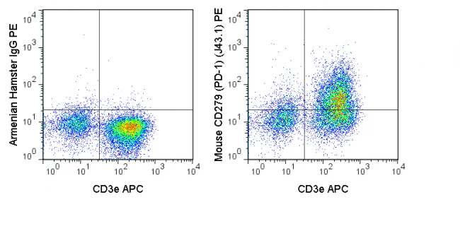
The J43.1 antibody is specific for mouse CD279, also known as programmed death-1 (PD-1), a 55 kDa glycoprotein which can co-regulate T cell antigen receptor signaling and therefore modulate T cell activation. PD-1 exists in a monomeric form that is expressed by CD4- CD8- thymocytes, where it participates in the processes of clonal selection, elimination of autoreactive lymphocytes, and development of tolerance. PD-1 expression is also inducible upon activation of mature T cells, where it has been proposed to interact with the co-stimulatory receptor CD80 to limit T cell activation. Two ligands for PD-1, known as PD-L1 (B7-H1) and PD-L2 (B7-DC) are differentially expressed on T and B cells, monocytes, macrophages, NK cells or dendritic cells. PD-1 is a member of a family of receptors including CD28 and CTLA-4 (CD152), which interact with “B7” ligands to provide a balance of co-stimulatory /co-inhibitory signaling important in T cell activation, tolerance, and autoimmunity.
The J43.1 antibody may be used as a marker for PD-1 expression, and is commonly used for analysis of receptor-ligand interaction and function(s) in vitro and in vivo. Please choose the appropriate format for each application.
| Name | PE Anti-Mouse CD279 (PD-1) (J43.1) |
|---|---|
| Cat. No. | 50-9985 |
| Alternative Names | PDCD1, PD1, SLEB2 , PD-1 |
| Gene ID | 18566 |
| Clone | J43.1 |
| Isotype | Armenian Hamster IgG |
| Reactivity | Mouse |
| Format | PE |
| Application | Flow Cytometry |
PE Anti-Mouse CD279 (PD-1) (J43.1)
The J43.1 antibody is specific for mouse CD279, also known as programmed death-1 (PD-1), a 55 kDa glycoprotein which can co-regulate T cell antigen receptor signaling and therefore modulate T cell activation. PD-1 exists in a monomeric form that is expressed by CD4- CD8- thymocytes, where it participates in the processes of clonal selection, elimination of autoreactive lymphocytes, and development of tolerance. PD-1 expression is also inducible upon activation of mature T cells, where it has been proposed to interact with the co-stimulatory receptor CD80 to limit T cell activation. Two ligands for PD-1, known as PD-L1 (B7-H1) and PD-L2 (B7-DC) are differentially expressed on T and B cells, monocytes, macrophages, NK cells or dendritic cells. PD-1 is a member of a family of receptors including CD28 and CTLA-4 (CD152), which interact with “B7” ligands to provide a balance of co-stimulatory /co-inhibitory signaling important in T cell activation, tolerance, and autoimmunity.
The J43.1 antibody may be used as a marker for PD-1 expression, and is commonly used for analysis of receptor-ligand interaction and function(s) in vitro and in vivo. Please choose the appropriate format for each application.
| Name | PE Anti-Mouse CD279 (PD-1) (J43.1) |
|---|---|
| Cat. No. | 50-9985 |
| Alternative Names | PDCD1, PD1, SLEB2 , PD-1 |
| Gene ID | 18566 |
| Clone | J43.1 |
| Isotype | Armenian Hamster IgG |
| Reactivity | Mouse |
| Format | PE |
| Application | Flow Cytometry |
Product Information
Product Information
Shipping & Returns
Shipping & Returns
Description
The J43.1 antibody is specific for mouse CD279, also known as programmed death-1 (PD-1), a 55 kDa glycoprotein which can co-regulate T cell antigen receptor signaling and therefore modulate T cell activation. PD-1 exists in a monomeric form that is expressed by CD4- CD8- thymocytes, where it participates in the processes of clonal selection, elimination of autoreactive lymphocytes, and development of tolerance. PD-1 expression is also inducible upon activation of mature T cells, where it has been proposed to interact with the co-stimulatory receptor CD80 to limit T cell activation. Two ligands for PD-1, known as PD-L1 (B7-H1) and PD-L2 (B7-DC) are differentially expressed on T and B cells, monocytes, macrophages, NK cells or dendritic cells. PD-1 is a member of a family of receptors including CD28 and CTLA-4 (CD152), which interact with “B7” ligands to provide a balance of co-stimulatory /co-inhibitory signaling important in T cell activation, tolerance, and autoimmunity.
The J43.1 antibody may be used as a marker for PD-1 expression, and is commonly used for analysis of receptor-ligand interaction and function(s) in vitro and in vivo. Please choose the appropriate format for each application.
| Name | PE Anti-Mouse CD279 (PD-1) (J43.1) |
|---|---|
| Cat. No. | 50-9985 |
| Alternative Names | PDCD1, PD1, SLEB2 , PD-1 |
| Gene ID | 18566 |
| Clone | J43.1 |
| Isotype | Armenian Hamster IgG |
| Reactivity | Mouse |
| Format | PE |
| Application | Flow Cytometry |