TRA-1-60 (Podocalyxin) Monoclonal Antibody (TRA-1-60), PE, eBioscience
PRODUCT DETAILS
Host: Mouse
Isotype: IgM
Clonality: Monoclonal
Clone: TRA-1-60
Format: PE
Reactivity: Hu
Application: Flow Cytometry
Tested Dilution: 1 µg/test
Concentration: 0.2 mg/mL
Storage: 4°C, store in dark, DO NOT FREEZE!
Formulation: PBS with 0.09% sodium azide; pH 7.2
Purification: Affinity chromatography
Data Sheet: TDS
Specific Information
Description: The TRA-1-60 antibody recognizes a protein expressed on undifferentiated human embryonic stem cells (ES), embyronal carcinoma cells (EC), and embryonic germ cells (EG). Like other stem cell specific markers, the epitope recognized by the TRA-1-60 antibody is lost upon cell differentiation. Contrary to early reports that the TRA-1-60 epitope can be destroyed by neuraminidase digestion, new strudies have shown that TRA-1-60 recognizes a neurominidase resistent antigen. The TRA-1-60 antibody is known to specifically recognize a carbohydrate epitope on a keratan sulfated glycoprotein recently identified as podocalyxin, a member of the CD34-related family of sialomucins. Podocalyxin is a transmembrane glycoprotein originally identified on epithelial glomerular cells known as podocytes, and the protein has also been implicated in the development of aggressiveness in a variety of cancers, including breast and prostate cancer.
Applications Reported: This TRA-1-60 antibody has been reported for use in flow cytometric analysis.
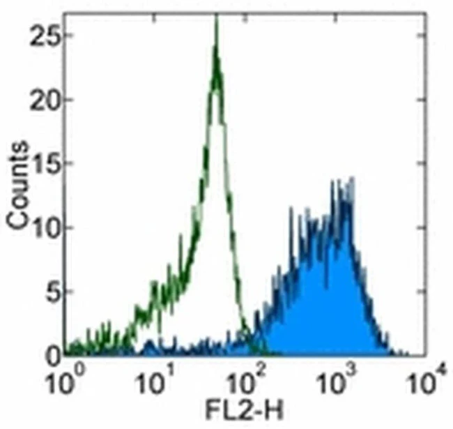
Applications Tested: This TRA-1-60 antibody has been tested by flow cytometric analysis of the human embryonal carcinoma (EC) line 2102Ep. This can be used at less than or equal to 1 µg per test. A test is defined as the amount (µg) of antibody that will stain a cell sample in a final volume of 100 µL. Cell number should be determined empirically but can range from 10^5 to 10^8 cells/test. It is recommended that the antibody be carefully titrated for optimal performance in the assay of interest.
Excitation: 488-561 nm; Emission: 578 nm; Laser: Blue Laser, Green Laser, Yellow-Green Laser.
Filtration: 0.2 µm post-manufacturing filtered.
For Research Use Only. Not for use in diagnostic procedures. Not for resale without express authorization.
Original: $281.00
-70%$281.00
$84.30TRA-1-60 (Podocalyxin) Monoclonal Antibody (TRA-1-60), PE, eBioscience
PRODUCT DETAILS
Host: Mouse
Isotype: IgM
Clonality: Monoclonal
Clone: TRA-1-60
Format: PE
Reactivity: Hu
Application: Flow Cytometry
Tested Dilution: 1 µg/test
Concentration: 0.2 mg/mL
Storage: 4°C, store in dark, DO NOT FREEZE!
Formulation: PBS with 0.09% sodium azide; pH 7.2
Purification: Affinity chromatography
Data Sheet: TDS
Specific Information
Description: The TRA-1-60 antibody recognizes a protein expressed on undifferentiated human embryonic stem cells (ES), embyronal carcinoma cells (EC), and embryonic germ cells (EG). Like other stem cell specific markers, the epitope recognized by the TRA-1-60 antibody is lost upon cell differentiation. Contrary to early reports that the TRA-1-60 epitope can be destroyed by neuraminidase digestion, new strudies have shown that TRA-1-60 recognizes a neurominidase resistent antigen. The TRA-1-60 antibody is known to specifically recognize a carbohydrate epitope on a keratan sulfated glycoprotein recently identified as podocalyxin, a member of the CD34-related family of sialomucins. Podocalyxin is a transmembrane glycoprotein originally identified on epithelial glomerular cells known as podocytes, and the protein has also been implicated in the development of aggressiveness in a variety of cancers, including breast and prostate cancer.
Applications Reported: This TRA-1-60 antibody has been reported for use in flow cytometric analysis.
Applications Tested: This TRA-1-60 antibody has been tested by flow cytometric analysis of the human embryonal carcinoma (EC) line 2102Ep. This can be used at less than or equal to 1 µg per test. A test is defined as the amount (µg) of antibody that will stain a cell sample in a final volume of 100 µL. Cell number should be determined empirically but can range from 10^5 to 10^8 cells/test. It is recommended that the antibody be carefully titrated for optimal performance in the assay of interest.
Excitation: 488-561 nm; Emission: 578 nm; Laser: Blue Laser, Green Laser, Yellow-Green Laser.
Filtration: 0.2 µm post-manufacturing filtered.
For Research Use Only. Not for use in diagnostic procedures. Not for resale without express authorization.
Product Information
Product Information
Shipping & Returns
Shipping & Returns
Description
PRODUCT DETAILS
Host: Mouse
Isotype: IgM
Clonality: Monoclonal
Clone: TRA-1-60
Format: PE
Reactivity: Hu
Application: Flow Cytometry
Tested Dilution: 1 µg/test
Concentration: 0.2 mg/mL
Storage: 4°C, store in dark, DO NOT FREEZE!
Formulation: PBS with 0.09% sodium azide; pH 7.2
Purification: Affinity chromatography
Data Sheet: TDS
Specific Information
Description: The TRA-1-60 antibody recognizes a protein expressed on undifferentiated human embryonic stem cells (ES), embyronal carcinoma cells (EC), and embryonic germ cells (EG). Like other stem cell specific markers, the epitope recognized by the TRA-1-60 antibody is lost upon cell differentiation. Contrary to early reports that the TRA-1-60 epitope can be destroyed by neuraminidase digestion, new strudies have shown that TRA-1-60 recognizes a neurominidase resistent antigen. The TRA-1-60 antibody is known to specifically recognize a carbohydrate epitope on a keratan sulfated glycoprotein recently identified as podocalyxin, a member of the CD34-related family of sialomucins. Podocalyxin is a transmembrane glycoprotein originally identified on epithelial glomerular cells known as podocytes, and the protein has also been implicated in the development of aggressiveness in a variety of cancers, including breast and prostate cancer.
Applications Reported: This TRA-1-60 antibody has been reported for use in flow cytometric analysis.
Applications Tested: This TRA-1-60 antibody has been tested by flow cytometric analysis of the human embryonal carcinoma (EC) line 2102Ep. This can be used at less than or equal to 1 µg per test. A test is defined as the amount (µg) of antibody that will stain a cell sample in a final volume of 100 µL. Cell number should be determined empirically but can range from 10^5 to 10^8 cells/test. It is recommended that the antibody be carefully titrated for optimal performance in the assay of interest.
Excitation: 488-561 nm; Emission: 578 nm; Laser: Blue Laser, Green Laser, Yellow-Green Laser.
Filtration: 0.2 µm post-manufacturing filtered.
For Research Use Only. Not for use in diagnostic procedures. Not for resale without express authorization.


