TER-119 Monoclonal Antibody (TER-119), PE-Cyanine5, eBioscience
PRODUCT DETAILS
Host: Rat
Isotype: IgG2b, kappa
Clonality: Monoclonal
Clone: TER-119
Format: PE-Cyanine5
Reactivity: Ms
Application: Flow Cytometry
Tested Dilution: 0.25 µg/test
Concentration: 0.2 mg/mL
Storage: 4°C, store in dark, DO NOT FREEZE!
Formulation: PBS with 0.09% sodium azide; pH 7.2
Purification: Affinity chromatography
Data Sheet: TDS
Specific Information
Description: The TER-119 monoclonal antibody reacts with mouse erythroid cells from early proerythroblast to mature erythrocyte stages. The TER-119 antigen is present in yolk sac, fetal and newborn liver, but is not expressed by cells carrying BFU-E and CFU-E activities. Several erythroleukemia cell lines tested so far are negative for expression of TER-119 antigen even after dimethylsulfoxide stimulation. Biochemical and molecular analysis of the TER-119 antigen indicate that this molecule is associated with the surface glycophorin A, but is not a typical glycophorin.
Applications Reported: The TER-119 antibody has been reported for use in flow cytometric analysis.
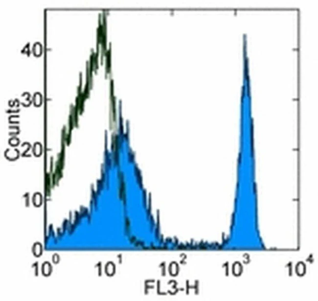
Applications Tested: The TER-119 antibody has been tested by flow cytometric analysis of mouse splenocytes and bone marrow cells. This can be used at less than or equal to 0.25 µg per test. A test is defined as the amount (µg) of antibody that will stain a cell sample in a final volume of 100 µL. Cell number should be determined empirically but can range from 10^5 to 10^8 cells/test. It is recommended that the antibody be carefully titrated for optimal performance in the assay of interest.
Light sensitivity: This tandem dye is sensitive photo-induced oxidation. Please protect this vial and stained samples from light.
Fixation: Samples can be stored in IC Fixation Buffer (Product # 00-822-49) (100 µL cell sample + 100 µL IC Fixation Buffer) or 1-step Fix/Lyse Solution (Product # 00-5333-54) for up to 3 days in the dark at 4°C with minimal impact on brightness and FRET efficiency/compensation. Some generalizations regarding fluorophore performance after fixation can be made, but clone specific performance should be determined empirically.
Excitation: 488-561 nm; Emission: 667 nm; Laser: Blue Laser, Green Laser, Yellow-Green Laser.
Filtration: 0.2 µm post-manufacturing filtered.
For Research Use Only. Not for use in diagnostic procedures. Not for resale without express authorization.
TER-119 Monoclonal Antibody (TER-119), PE-Cyanine5, eBioscience
PRODUCT DETAILS
Host: Rat
Isotype: IgG2b, kappa
Clonality: Monoclonal
Clone: TER-119
Format: PE-Cyanine5
Reactivity: Ms
Application: Flow Cytometry
Tested Dilution: 0.25 µg/test
Concentration: 0.2 mg/mL
Storage: 4°C, store in dark, DO NOT FREEZE!
Formulation: PBS with 0.09% sodium azide; pH 7.2
Purification: Affinity chromatography
Data Sheet: TDS
Specific Information
Description: The TER-119 monoclonal antibody reacts with mouse erythroid cells from early proerythroblast to mature erythrocyte stages. The TER-119 antigen is present in yolk sac, fetal and newborn liver, but is not expressed by cells carrying BFU-E and CFU-E activities. Several erythroleukemia cell lines tested so far are negative for expression of TER-119 antigen even after dimethylsulfoxide stimulation. Biochemical and molecular analysis of the TER-119 antigen indicate that this molecule is associated with the surface glycophorin A, but is not a typical glycophorin.
Applications Reported: The TER-119 antibody has been reported for use in flow cytometric analysis.
Applications Tested: The TER-119 antibody has been tested by flow cytometric analysis of mouse splenocytes and bone marrow cells. This can be used at less than or equal to 0.25 µg per test. A test is defined as the amount (µg) of antibody that will stain a cell sample in a final volume of 100 µL. Cell number should be determined empirically but can range from 10^5 to 10^8 cells/test. It is recommended that the antibody be carefully titrated for optimal performance in the assay of interest.
Light sensitivity: This tandem dye is sensitive photo-induced oxidation. Please protect this vial and stained samples from light.
Fixation: Samples can be stored in IC Fixation Buffer (Product # 00-822-49) (100 µL cell sample + 100 µL IC Fixation Buffer) or 1-step Fix/Lyse Solution (Product # 00-5333-54) for up to 3 days in the dark at 4°C with minimal impact on brightness and FRET efficiency/compensation. Some generalizations regarding fluorophore performance after fixation can be made, but clone specific performance should be determined empirically.
Excitation: 488-561 nm; Emission: 667 nm; Laser: Blue Laser, Green Laser, Yellow-Green Laser.
Filtration: 0.2 µm post-manufacturing filtered.
For Research Use Only. Not for use in diagnostic procedures. Not for resale without express authorization.
Product Information
Product Information
Shipping & Returns
Shipping & Returns
Description
PRODUCT DETAILS
Host: Rat
Isotype: IgG2b, kappa
Clonality: Monoclonal
Clone: TER-119
Format: PE-Cyanine5
Reactivity: Ms
Application: Flow Cytometry
Tested Dilution: 0.25 µg/test
Concentration: 0.2 mg/mL
Storage: 4°C, store in dark, DO NOT FREEZE!
Formulation: PBS with 0.09% sodium azide; pH 7.2
Purification: Affinity chromatography
Data Sheet: TDS
Specific Information
Description: The TER-119 monoclonal antibody reacts with mouse erythroid cells from early proerythroblast to mature erythrocyte stages. The TER-119 antigen is present in yolk sac, fetal and newborn liver, but is not expressed by cells carrying BFU-E and CFU-E activities. Several erythroleukemia cell lines tested so far are negative for expression of TER-119 antigen even after dimethylsulfoxide stimulation. Biochemical and molecular analysis of the TER-119 antigen indicate that this molecule is associated with the surface glycophorin A, but is not a typical glycophorin.
Applications Reported: The TER-119 antibody has been reported for use in flow cytometric analysis.
Applications Tested: The TER-119 antibody has been tested by flow cytometric analysis of mouse splenocytes and bone marrow cells. This can be used at less than or equal to 0.25 µg per test. A test is defined as the amount (µg) of antibody that will stain a cell sample in a final volume of 100 µL. Cell number should be determined empirically but can range from 10^5 to 10^8 cells/test. It is recommended that the antibody be carefully titrated for optimal performance in the assay of interest.
Light sensitivity: This tandem dye is sensitive photo-induced oxidation. Please protect this vial and stained samples from light.
Fixation: Samples can be stored in IC Fixation Buffer (Product # 00-822-49) (100 µL cell sample + 100 µL IC Fixation Buffer) or 1-step Fix/Lyse Solution (Product # 00-5333-54) for up to 3 days in the dark at 4°C with minimal impact on brightness and FRET efficiency/compensation. Some generalizations regarding fluorophore performance after fixation can be made, but clone specific performance should be determined empirically.
Excitation: 488-561 nm; Emission: 667 nm; Laser: Blue Laser, Green Laser, Yellow-Green Laser.
Filtration: 0.2 µm post-manufacturing filtered.
For Research Use Only. Not for use in diagnostic procedures. Not for resale without express authorization.


