Tau Monoclonal Antibody (HT7), Alexa Fluor 647, eBioscience
PRODUCT DETAILS
Host: Mouse
Isotype: IgG1, kappa
Clonality: Monoclonal
Clone: HT7
Format: Alexa Fluor 647
Reactivity: Hu
Application: Flow Cytometry
Tested Dilution: 5 µL (0.25 µg)/test
Concentration: 5 μL/Test
Storage: 4°C, store in dark, DO NOT FREEZE!
Formulation: PBS with BSA and 0.09% sodium azide; pH 7.2
Purification: Affinity chromatography
Data Sheet: TDS
Specific Information
Description: This HT7 monoclonal antibody recognizes human and bovine Tau protein. The epitope of this antibody has been mapped to residues 159 through 163, corresponding to the amino acid sequence PPGQK of human Tau40. In certain cell lines and tissue other than brain, clone HT7 may show a low but distinct amount of nonspecific staining. Therefore we recommend the use
of clone HT7 to detect Tau expression by flow cytometry in human Tau transfected cells or tissue derived from human Tau transgenic mice. This antibody does not cross-react with murine Tau.
Applications Reported: This HT7 antibody has been reported for use in flow cytometric analysis.
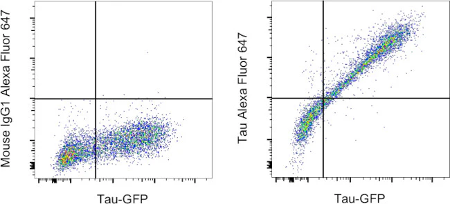
Applications Tested: This HT7 antibody has been pre-diluted and tested by flow cytometric analysis of Human Tau transfected 293 HEK cells using the Intracellular Fixation & Permeabilization Buffer Set (Product # 88-8824-00) and protocol. Please refer to "Staining Intracellular Antigens for Flow Cytometry, Protocol A: Two step protocol for intracellular (cytoplasmic) proteins" located at www.thermofisher.com/flowprotocols . This antibody may be used at 5 µL (0.25 µg) per test. A test is defined as the amount (µg) of antibody that will stain a cell sample in a final volume of 100 µL. Cell number should be determined empirically but can range from 10^5 to 10^8 cells/test.
Excitation: 633-647 nm; Emission: 668 nm; Laser: Red Laser.
For Research Use Only. Not for use in diagnostic procedures. Not for resale without express authorization.
Original: $568.00
-70%$568.00
$170.40Tau Monoclonal Antibody (HT7), Alexa Fluor 647, eBioscience
PRODUCT DETAILS
Host: Mouse
Isotype: IgG1, kappa
Clonality: Monoclonal
Clone: HT7
Format: Alexa Fluor 647
Reactivity: Hu
Application: Flow Cytometry
Tested Dilution: 5 µL (0.25 µg)/test
Concentration: 5 μL/Test
Storage: 4°C, store in dark, DO NOT FREEZE!
Formulation: PBS with BSA and 0.09% sodium azide; pH 7.2
Purification: Affinity chromatography
Data Sheet: TDS
Specific Information
Description: This HT7 monoclonal antibody recognizes human and bovine Tau protein. The epitope of this antibody has been mapped to residues 159 through 163, corresponding to the amino acid sequence PPGQK of human Tau40. In certain cell lines and tissue other than brain, clone HT7 may show a low but distinct amount of nonspecific staining. Therefore we recommend the use
of clone HT7 to detect Tau expression by flow cytometry in human Tau transfected cells or tissue derived from human Tau transgenic mice. This antibody does not cross-react with murine Tau.
Applications Reported: This HT7 antibody has been reported for use in flow cytometric analysis.
Applications Tested: This HT7 antibody has been pre-diluted and tested by flow cytometric analysis of Human Tau transfected 293 HEK cells using the Intracellular Fixation & Permeabilization Buffer Set (Product # 88-8824-00) and protocol. Please refer to "Staining Intracellular Antigens for Flow Cytometry, Protocol A: Two step protocol for intracellular (cytoplasmic) proteins" located at www.thermofisher.com/flowprotocols . This antibody may be used at 5 µL (0.25 µg) per test. A test is defined as the amount (µg) of antibody that will stain a cell sample in a final volume of 100 µL. Cell number should be determined empirically but can range from 10^5 to 10^8 cells/test.
Excitation: 633-647 nm; Emission: 668 nm; Laser: Red Laser.
For Research Use Only. Not for use in diagnostic procedures. Not for resale without express authorization.
Product Information
Product Information
Shipping & Returns
Shipping & Returns
Description
PRODUCT DETAILS
Host: Mouse
Isotype: IgG1, kappa
Clonality: Monoclonal
Clone: HT7
Format: Alexa Fluor 647
Reactivity: Hu
Application: Flow Cytometry
Tested Dilution: 5 µL (0.25 µg)/test
Concentration: 5 μL/Test
Storage: 4°C, store in dark, DO NOT FREEZE!
Formulation: PBS with BSA and 0.09% sodium azide; pH 7.2
Purification: Affinity chromatography
Data Sheet: TDS
Specific Information
Description: This HT7 monoclonal antibody recognizes human and bovine Tau protein. The epitope of this antibody has been mapped to residues 159 through 163, corresponding to the amino acid sequence PPGQK of human Tau40. In certain cell lines and tissue other than brain, clone HT7 may show a low but distinct amount of nonspecific staining. Therefore we recommend the use
of clone HT7 to detect Tau expression by flow cytometry in human Tau transfected cells or tissue derived from human Tau transgenic mice. This antibody does not cross-react with murine Tau.
Applications Reported: This HT7 antibody has been reported for use in flow cytometric analysis.
Applications Tested: This HT7 antibody has been pre-diluted and tested by flow cytometric analysis of Human Tau transfected 293 HEK cells using the Intracellular Fixation & Permeabilization Buffer Set (Product # 88-8824-00) and protocol. Please refer to "Staining Intracellular Antigens for Flow Cytometry, Protocol A: Two step protocol for intracellular (cytoplasmic) proteins" located at www.thermofisher.com/flowprotocols . This antibody may be used at 5 µL (0.25 µg) per test. A test is defined as the amount (µg) of antibody that will stain a cell sample in a final volume of 100 µL. Cell number should be determined empirically but can range from 10^5 to 10^8 cells/test.
Excitation: 633-647 nm; Emission: 668 nm; Laser: Red Laser.
For Research Use Only. Not for use in diagnostic procedures. Not for resale without express authorization.


