Syk Monoclonal Antibody (4D10.1), PE, eBioscience
PRODUCT DETAILS
Host: Mouse
Isotype: IgG2a, kappa
Clonality: Monoclonal
Clone: 4D10.1
Format: PE
Reactivity: Hu
Application: Flow Cytometry
Tested Dilution: 5 µL (0.125 µg)/test
Concentration: 5 μL/Test
Storage: 4°C, store in dark, DO NOT FREEZE!
Formulation: PBS with BSA and 0.09% sodium azide; pH 7.2
Purification: Affinity chromatography
Data Sheet: TDS
Specific Information
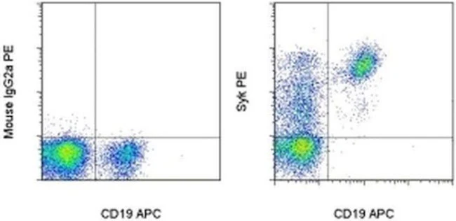
Description: This 4D10.1 monoclonal antibody recognizes human Syk, a 72-kDa member of the Syk/ZAP-70 family of non-receptor protein tyrosine kinases. Syk is expressed most highly in B cells, with lower expression in immature thymocytes, mast cells, and platelets. Syk can also be detected in fibroblasts, epithelial cells, hepatocytes, endothelial cells, and neuronal cells. This protein is a major component of signaling cascades downstream of the B and T cell antigen receptors and plays an essential role in lymphocyte development. Upon recruitment to immunoreceptor tyrosine-based activation motifs (ITAMs) on the antigen receptors, Syk is activated by phosphorylation on multiple tyrosines. Once activated, Syk phosphorylates proteins such as phospholipase C gamma and BLNK/SLP-65. Finally, abnormal Syk expression has been linked to tumor cell migration and invasion in several cancers.
Applications Reported: This 4D10.1 antibody has been reported for use in intracellular staining followed by flow cytometric analysis.
Applications Tested: This 4D10.1 antibody has been pre-titrated and tested by intracellular staining and flow cytometric analysis of normal human peripheral blood cells using the Foxp3/Transcription Factor Staining Buffer Set (Product # 00-5523-00) and protocol. Please refer to BestProtocols®: Protocol B: One step protocol for (nuclear) intracellular proteins located under the Resources Tab online. This can be used at 5 µL (0.125 µg) per test. A test is defined as the amount (µg) of antibody that will stain a cell sample in a final volume of 100 µL. Cell number should be determined empirically but can range from 10^5 to 10^8 cells/test.
Excitation: 488-561 nm; Emission: 578 nm; Laser: Blue Laser, Green Laser, Yellow-Green Laser.
Filtration: 0.2 µm post-manufacturing filtered.
For Research Use Only. Not for use in diagnostic procedures. Not for resale without express authorization.
Original: $412.00
-70%$412.00
$123.60Syk Monoclonal Antibody (4D10.1), PE, eBioscience
PRODUCT DETAILS
Host: Mouse
Isotype: IgG2a, kappa
Clonality: Monoclonal
Clone: 4D10.1
Format: PE
Reactivity: Hu
Application: Flow Cytometry
Tested Dilution: 5 µL (0.125 µg)/test
Concentration: 5 μL/Test
Storage: 4°C, store in dark, DO NOT FREEZE!
Formulation: PBS with BSA and 0.09% sodium azide; pH 7.2
Purification: Affinity chromatography
Data Sheet: TDS
Specific Information
Description: This 4D10.1 monoclonal antibody recognizes human Syk, a 72-kDa member of the Syk/ZAP-70 family of non-receptor protein tyrosine kinases. Syk is expressed most highly in B cells, with lower expression in immature thymocytes, mast cells, and platelets. Syk can also be detected in fibroblasts, epithelial cells, hepatocytes, endothelial cells, and neuronal cells. This protein is a major component of signaling cascades downstream of the B and T cell antigen receptors and plays an essential role in lymphocyte development. Upon recruitment to immunoreceptor tyrosine-based activation motifs (ITAMs) on the antigen receptors, Syk is activated by phosphorylation on multiple tyrosines. Once activated, Syk phosphorylates proteins such as phospholipase C gamma and BLNK/SLP-65. Finally, abnormal Syk expression has been linked to tumor cell migration and invasion in several cancers.
Applications Reported: This 4D10.1 antibody has been reported for use in intracellular staining followed by flow cytometric analysis.
Applications Tested: This 4D10.1 antibody has been pre-titrated and tested by intracellular staining and flow cytometric analysis of normal human peripheral blood cells using the Foxp3/Transcription Factor Staining Buffer Set (Product # 00-5523-00) and protocol. Please refer to BestProtocols®: Protocol B: One step protocol for (nuclear) intracellular proteins located under the Resources Tab online. This can be used at 5 µL (0.125 µg) per test. A test is defined as the amount (µg) of antibody that will stain a cell sample in a final volume of 100 µL. Cell number should be determined empirically but can range from 10^5 to 10^8 cells/test.
Excitation: 488-561 nm; Emission: 578 nm; Laser: Blue Laser, Green Laser, Yellow-Green Laser.
Filtration: 0.2 µm post-manufacturing filtered.
For Research Use Only. Not for use in diagnostic procedures. Not for resale without express authorization.
Product Information
Product Information
Shipping & Returns
Shipping & Returns
Description
PRODUCT DETAILS
Host: Mouse
Isotype: IgG2a, kappa
Clonality: Monoclonal
Clone: 4D10.1
Format: PE
Reactivity: Hu
Application: Flow Cytometry
Tested Dilution: 5 µL (0.125 µg)/test
Concentration: 5 μL/Test
Storage: 4°C, store in dark, DO NOT FREEZE!
Formulation: PBS with BSA and 0.09% sodium azide; pH 7.2
Purification: Affinity chromatography
Data Sheet: TDS
Specific Information
Description: This 4D10.1 monoclonal antibody recognizes human Syk, a 72-kDa member of the Syk/ZAP-70 family of non-receptor protein tyrosine kinases. Syk is expressed most highly in B cells, with lower expression in immature thymocytes, mast cells, and platelets. Syk can also be detected in fibroblasts, epithelial cells, hepatocytes, endothelial cells, and neuronal cells. This protein is a major component of signaling cascades downstream of the B and T cell antigen receptors and plays an essential role in lymphocyte development. Upon recruitment to immunoreceptor tyrosine-based activation motifs (ITAMs) on the antigen receptors, Syk is activated by phosphorylation on multiple tyrosines. Once activated, Syk phosphorylates proteins such as phospholipase C gamma and BLNK/SLP-65. Finally, abnormal Syk expression has been linked to tumor cell migration and invasion in several cancers.
Applications Reported: This 4D10.1 antibody has been reported for use in intracellular staining followed by flow cytometric analysis.
Applications Tested: This 4D10.1 antibody has been pre-titrated and tested by intracellular staining and flow cytometric analysis of normal human peripheral blood cells using the Foxp3/Transcription Factor Staining Buffer Set (Product # 00-5523-00) and protocol. Please refer to BestProtocols®: Protocol B: One step protocol for (nuclear) intracellular proteins located under the Resources Tab online. This can be used at 5 µL (0.125 µg) per test. A test is defined as the amount (µg) of antibody that will stain a cell sample in a final volume of 100 µL. Cell number should be determined empirically but can range from 10^5 to 10^8 cells/test.
Excitation: 488-561 nm; Emission: 578 nm; Laser: Blue Laser, Green Laser, Yellow-Green Laser.
Filtration: 0.2 µm post-manufacturing filtered.
For Research Use Only. Not for use in diagnostic procedures. Not for resale without express authorization.


