Siglec-G Monoclonal Antibody (SH2.1), PerCP-eFluor 710, eBioscience
PRODUCT DETAILS
Host: Mouse
Isotype: IgG1, kappa
Clonality: Monoclonal
Clone: SH2.1
Format: PerCP-eFluor 710
Reactivity: Ms
Application: Flow Cytometry
Tested Dilution: 0.125 µg/test
Concentration: 0.2 mg/mL
Storage: 4°C, store in dark, DO NOT FREEZE!
Formulation: PBS with 0.09% sodium azide; pH 7.2
Purification: Affinity chromatography
Data Sheet: TDS
Specific Information
Description: The SH2.1 monoclonal antibody reacts with mouse siglec-G, which is also known as sialic acid-binding immunoglobulin-like lectin-G. Siglec-G is an inhibitory receptor that is expressed in a B cell-specific manner with B1 B cells expressing the highest levels of siglec-G. When overexpressed, siglec-G inhibits B cell receptor-mediated calcium signaling. Siglec-G-deficient mice show a massive expansion of the B1a cell population as well as higher titers of natural IgM antibodies. Siglec-G-deficient B1a cells show a lower level of spontaneous apoptosis and a prolonged life span, which may result from higher expression levels of the transcription factor NFATc1. Siglec-G-dependent negative regulation exists in B1 cells, which may explain the naturally muted signaling response of B1 cells.
Applications Reported: This SH2.1 antibody has been reported for use in flow cytometric analysis.
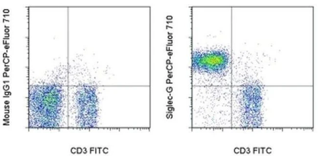
Applications Tested: This SH2.1 antibody has been tested by flow cytometric analysis of mouse splenocytes. This can be used at less than or equal to 0.125 µg per test. A test is defined as the amount (µg) of antibody that will stain a cell sample in a final volume of 100 µL. Cell number should be determined empirically but can range from 10^5 to 10^8 cells/test. It is recommended that the antibody be carefully titrated for optimal performance in the assay of interest.
PerCP-eFluor® 710 emits at 710 nm and is excited with the blue laser (488 nm); it can be used in place of PerCP-Cyanine5.5. We recommend using a 710/50 bandpass filter, however, the 695/40 bandpass filter is an acceptable alternative. Please make sure that your instrument is capable of detecting this fluorochrome.
Fixation: Samples can be stored in IC Fixation Buffer (Product # 00-822-49) (100 µL cell sample + 100 µL IC Fixation Buffer) or 1-step Fix/Lyse Solution (Product # 00-5333-54) for up to 3 days in the dark at 4°C with minimal impact on brightness and FRET efficiency/compensation. Some generalizations regarding fluorophore performance after fixation can be made, but clone specific performance should be determined empirically.
Excitation: 488 nm; Emission: 710 nm; Laser: Blue Laser.
Filtration: 0.2 µm post-manufacturing filtered.
For Research Use Only. Not for use in diagnostic procedures. Not for resale without express authorization.
Siglec-G Monoclonal Antibody (SH2.1), PerCP-eFluor 710, eBioscience
PRODUCT DETAILS
Host: Mouse
Isotype: IgG1, kappa
Clonality: Monoclonal
Clone: SH2.1
Format: PerCP-eFluor 710
Reactivity: Ms
Application: Flow Cytometry
Tested Dilution: 0.125 µg/test
Concentration: 0.2 mg/mL
Storage: 4°C, store in dark, DO NOT FREEZE!
Formulation: PBS with 0.09% sodium azide; pH 7.2
Purification: Affinity chromatography
Data Sheet: TDS
Specific Information
Description: The SH2.1 monoclonal antibody reacts with mouse siglec-G, which is also known as sialic acid-binding immunoglobulin-like lectin-G. Siglec-G is an inhibitory receptor that is expressed in a B cell-specific manner with B1 B cells expressing the highest levels of siglec-G. When overexpressed, siglec-G inhibits B cell receptor-mediated calcium signaling. Siglec-G-deficient mice show a massive expansion of the B1a cell population as well as higher titers of natural IgM antibodies. Siglec-G-deficient B1a cells show a lower level of spontaneous apoptosis and a prolonged life span, which may result from higher expression levels of the transcription factor NFATc1. Siglec-G-dependent negative regulation exists in B1 cells, which may explain the naturally muted signaling response of B1 cells.
Applications Reported: This SH2.1 antibody has been reported for use in flow cytometric analysis.
Applications Tested: This SH2.1 antibody has been tested by flow cytometric analysis of mouse splenocytes. This can be used at less than or equal to 0.125 µg per test. A test is defined as the amount (µg) of antibody that will stain a cell sample in a final volume of 100 µL. Cell number should be determined empirically but can range from 10^5 to 10^8 cells/test. It is recommended that the antibody be carefully titrated for optimal performance in the assay of interest.
PerCP-eFluor® 710 emits at 710 nm and is excited with the blue laser (488 nm); it can be used in place of PerCP-Cyanine5.5. We recommend using a 710/50 bandpass filter, however, the 695/40 bandpass filter is an acceptable alternative. Please make sure that your instrument is capable of detecting this fluorochrome.
Fixation: Samples can be stored in IC Fixation Buffer (Product # 00-822-49) (100 µL cell sample + 100 µL IC Fixation Buffer) or 1-step Fix/Lyse Solution (Product # 00-5333-54) for up to 3 days in the dark at 4°C with minimal impact on brightness and FRET efficiency/compensation. Some generalizations regarding fluorophore performance after fixation can be made, but clone specific performance should be determined empirically.
Excitation: 488 nm; Emission: 710 nm; Laser: Blue Laser.
Filtration: 0.2 µm post-manufacturing filtered.
For Research Use Only. Not for use in diagnostic procedures. Not for resale without express authorization.
Product Information
Product Information
Shipping & Returns
Shipping & Returns
Description
PRODUCT DETAILS
Host: Mouse
Isotype: IgG1, kappa
Clonality: Monoclonal
Clone: SH2.1
Format: PerCP-eFluor 710
Reactivity: Ms
Application: Flow Cytometry
Tested Dilution: 0.125 µg/test
Concentration: 0.2 mg/mL
Storage: 4°C, store in dark, DO NOT FREEZE!
Formulation: PBS with 0.09% sodium azide; pH 7.2
Purification: Affinity chromatography
Data Sheet: TDS
Specific Information
Description: The SH2.1 monoclonal antibody reacts with mouse siglec-G, which is also known as sialic acid-binding immunoglobulin-like lectin-G. Siglec-G is an inhibitory receptor that is expressed in a B cell-specific manner with B1 B cells expressing the highest levels of siglec-G. When overexpressed, siglec-G inhibits B cell receptor-mediated calcium signaling. Siglec-G-deficient mice show a massive expansion of the B1a cell population as well as higher titers of natural IgM antibodies. Siglec-G-deficient B1a cells show a lower level of spontaneous apoptosis and a prolonged life span, which may result from higher expression levels of the transcription factor NFATc1. Siglec-G-dependent negative regulation exists in B1 cells, which may explain the naturally muted signaling response of B1 cells.
Applications Reported: This SH2.1 antibody has been reported for use in flow cytometric analysis.
Applications Tested: This SH2.1 antibody has been tested by flow cytometric analysis of mouse splenocytes. This can be used at less than or equal to 0.125 µg per test. A test is defined as the amount (µg) of antibody that will stain a cell sample in a final volume of 100 µL. Cell number should be determined empirically but can range from 10^5 to 10^8 cells/test. It is recommended that the antibody be carefully titrated for optimal performance in the assay of interest.
PerCP-eFluor® 710 emits at 710 nm and is excited with the blue laser (488 nm); it can be used in place of PerCP-Cyanine5.5. We recommend using a 710/50 bandpass filter, however, the 695/40 bandpass filter is an acceptable alternative. Please make sure that your instrument is capable of detecting this fluorochrome.
Fixation: Samples can be stored in IC Fixation Buffer (Product # 00-822-49) (100 µL cell sample + 100 µL IC Fixation Buffer) or 1-step Fix/Lyse Solution (Product # 00-5333-54) for up to 3 days in the dark at 4°C with minimal impact on brightness and FRET efficiency/compensation. Some generalizations regarding fluorophore performance after fixation can be made, but clone specific performance should be determined empirically.
Excitation: 488 nm; Emission: 710 nm; Laser: Blue Laser.
Filtration: 0.2 µm post-manufacturing filtered.
For Research Use Only. Not for use in diagnostic procedures. Not for resale without express authorization.


