Podoplanin Monoclonal Antibody (eBio8.1.1(8.1.1)), Super Bright 436, eBioscience
PRODUCT DETAILS
Host: Syrian Hamster
Isotype: IgG
Clonality: Monoclonal
Clone: eBio8.1.1(8.1.1)
Format: Super Bright 436
Reactivity: Ms
Application: Flow Cytometry
Tested Dilution: 0.25 µg/test
Concentration: 0.2 mg/mL
Storage: 4°C, store in dark, DO NOT FREEZE!
Formulation: PBS with BSA and 0.09% sodium azide; pH 7.2
Purification: Affinity chromatography
Data Sheet: TDS
Specific Information
Description: The 8.1.1 monoclonal antibody reacts with mouse podoplanin (T1a, gp38, aggrus), a 43 kDa transmembrane glycoprotein, named for its expression in kidney glomerular epithelial cells (podocytes). In addition, Podoplanin is expressed in epithelial and mesothelial cells such as intestinal epithelium, alveolar type I cells, podocytes, and mesothelium of the visceral peritoneum. It was also shown to be a potent marker for lymphatic endothelium. Podoplanin is also expressed by subcapsular epithelial cells of the murine thymus. Mice deficient in Podoplanin die at birth because of a respiratory defect and congenital lymphedema due to a failure in lymphatic pattern formation.
Applications Reported: This eBio8.1.1 (8.1.1) antibody has been reported for use in flow cytometric analysis.
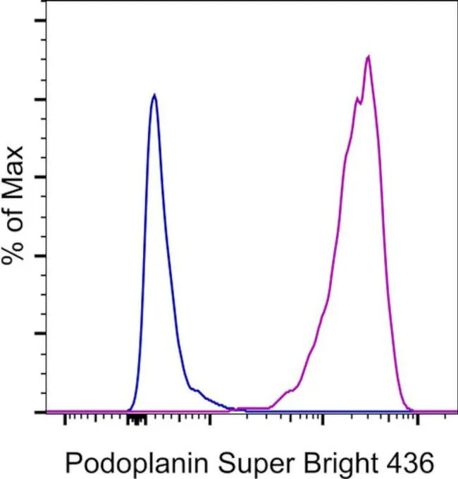
Applications Tested: This eBio8.1.1 (8.1.1) antibody has been tested by flow cytometric analysis of TE-71 cell line. This can be used at less than or equal to 0.25 µg per test. A test is defined as the amount (µg) of antibody that will stain a cell sample in a final volume of 100 µL. Cell number should be determined empirically but can range from 10^5 to 10^8 cells/test. It is recommended that the antibody be carefully titrated for optimal performance in the assay of interest.
Super Bright 436 can be excited with the violet laser line (405 nm) and emits at 436 nm. We recommend using a 450/50 bandpass filter, or equivalent. Please make sure that your instrument is capable of detecting this fluorochrome.
When using two or more Super Bright dye-conjugated antibodies in a staining panel, it is recommended to use Super Bright Complete Staining Buffer (Product # SB-4401) to minimize any non-specific polymer interactions. Please refer to the datasheet for Super Bright Staining Buffer for more information.
Fixation: Samples can be stored in IC Fixation Buffer (Product # 00-8222) (100 µL of cell sample + 100 µL of IC Fixation Buffer) or 1-step Fix/Lyse Solution (Product # 00-5333) for up to 3 days in the dark at 4°C with minimal impact on brightness and FRET efficiency/compensation. Some generalizations regarding fluorophore performance after fixation can be made, but clone specific performance should be determined empirically.
Excitation: 405 nm; Emission: 436 nm; Laser: Violet Laser
Super Bright Polymer Dyes are sold under license from Becton, Dickinson and Company.
For Research Use Only. Not for use in diagnostic procedures. Not for resale without express authorization.
Podoplanin Monoclonal Antibody (eBio8.1.1(8.1.1)), Super Bright 436, eBioscience
PRODUCT DETAILS
Host: Syrian Hamster
Isotype: IgG
Clonality: Monoclonal
Clone: eBio8.1.1(8.1.1)
Format: Super Bright 436
Reactivity: Ms
Application: Flow Cytometry
Tested Dilution: 0.25 µg/test
Concentration: 0.2 mg/mL
Storage: 4°C, store in dark, DO NOT FREEZE!
Formulation: PBS with BSA and 0.09% sodium azide; pH 7.2
Purification: Affinity chromatography
Data Sheet: TDS
Specific Information
Description: The 8.1.1 monoclonal antibody reacts with mouse podoplanin (T1a, gp38, aggrus), a 43 kDa transmembrane glycoprotein, named for its expression in kidney glomerular epithelial cells (podocytes). In addition, Podoplanin is expressed in epithelial and mesothelial cells such as intestinal epithelium, alveolar type I cells, podocytes, and mesothelium of the visceral peritoneum. It was also shown to be a potent marker for lymphatic endothelium. Podoplanin is also expressed by subcapsular epithelial cells of the murine thymus. Mice deficient in Podoplanin die at birth because of a respiratory defect and congenital lymphedema due to a failure in lymphatic pattern formation.
Applications Reported: This eBio8.1.1 (8.1.1) antibody has been reported for use in flow cytometric analysis.
Applications Tested: This eBio8.1.1 (8.1.1) antibody has been tested by flow cytometric analysis of TE-71 cell line. This can be used at less than or equal to 0.25 µg per test. A test is defined as the amount (µg) of antibody that will stain a cell sample in a final volume of 100 µL. Cell number should be determined empirically but can range from 10^5 to 10^8 cells/test. It is recommended that the antibody be carefully titrated for optimal performance in the assay of interest.
Super Bright 436 can be excited with the violet laser line (405 nm) and emits at 436 nm. We recommend using a 450/50 bandpass filter, or equivalent. Please make sure that your instrument is capable of detecting this fluorochrome.
When using two or more Super Bright dye-conjugated antibodies in a staining panel, it is recommended to use Super Bright Complete Staining Buffer (Product # SB-4401) to minimize any non-specific polymer interactions. Please refer to the datasheet for Super Bright Staining Buffer for more information.
Fixation: Samples can be stored in IC Fixation Buffer (Product # 00-8222) (100 µL of cell sample + 100 µL of IC Fixation Buffer) or 1-step Fix/Lyse Solution (Product # 00-5333) for up to 3 days in the dark at 4°C with minimal impact on brightness and FRET efficiency/compensation. Some generalizations regarding fluorophore performance after fixation can be made, but clone specific performance should be determined empirically.
Excitation: 405 nm; Emission: 436 nm; Laser: Violet Laser
Super Bright Polymer Dyes are sold under license from Becton, Dickinson and Company.
For Research Use Only. Not for use in diagnostic procedures. Not for resale without express authorization.
Product Information
Product Information
Shipping & Returns
Shipping & Returns
Description
PRODUCT DETAILS
Host: Syrian Hamster
Isotype: IgG
Clonality: Monoclonal
Clone: eBio8.1.1(8.1.1)
Format: Super Bright 436
Reactivity: Ms
Application: Flow Cytometry
Tested Dilution: 0.25 µg/test
Concentration: 0.2 mg/mL
Storage: 4°C, store in dark, DO NOT FREEZE!
Formulation: PBS with BSA and 0.09% sodium azide; pH 7.2
Purification: Affinity chromatography
Data Sheet: TDS
Specific Information
Description: The 8.1.1 monoclonal antibody reacts with mouse podoplanin (T1a, gp38, aggrus), a 43 kDa transmembrane glycoprotein, named for its expression in kidney glomerular epithelial cells (podocytes). In addition, Podoplanin is expressed in epithelial and mesothelial cells such as intestinal epithelium, alveolar type I cells, podocytes, and mesothelium of the visceral peritoneum. It was also shown to be a potent marker for lymphatic endothelium. Podoplanin is also expressed by subcapsular epithelial cells of the murine thymus. Mice deficient in Podoplanin die at birth because of a respiratory defect and congenital lymphedema due to a failure in lymphatic pattern formation.
Applications Reported: This eBio8.1.1 (8.1.1) antibody has been reported for use in flow cytometric analysis.
Applications Tested: This eBio8.1.1 (8.1.1) antibody has been tested by flow cytometric analysis of TE-71 cell line. This can be used at less than or equal to 0.25 µg per test. A test is defined as the amount (µg) of antibody that will stain a cell sample in a final volume of 100 µL. Cell number should be determined empirically but can range from 10^5 to 10^8 cells/test. It is recommended that the antibody be carefully titrated for optimal performance in the assay of interest.
Super Bright 436 can be excited with the violet laser line (405 nm) and emits at 436 nm. We recommend using a 450/50 bandpass filter, or equivalent. Please make sure that your instrument is capable of detecting this fluorochrome.
When using two or more Super Bright dye-conjugated antibodies in a staining panel, it is recommended to use Super Bright Complete Staining Buffer (Product # SB-4401) to minimize any non-specific polymer interactions. Please refer to the datasheet for Super Bright Staining Buffer for more information.
Fixation: Samples can be stored in IC Fixation Buffer (Product # 00-8222) (100 µL of cell sample + 100 µL of IC Fixation Buffer) or 1-step Fix/Lyse Solution (Product # 00-5333) for up to 3 days in the dark at 4°C with minimal impact on brightness and FRET efficiency/compensation. Some generalizations regarding fluorophore performance after fixation can be made, but clone specific performance should be determined empirically.
Excitation: 405 nm; Emission: 436 nm; Laser: Violet Laser
Super Bright Polymer Dyes are sold under license from Becton, Dickinson and Company.
For Research Use Only. Not for use in diagnostic procedures. Not for resale without express authorization.


