PLZF Monoclonal Antibody (Mags.21F7), Brilliant Violet 711, eBioscience
PRODUCT DETAILS
Host: Mouse
Isotype: IgG1, kappa
Clonality: Monoclonal
Clone: Mags.21F7
Format: Brilliant Violet 711
Reactivity: Hu, Ms
Application: Flow Cytometry
Tested Dilution: 1.0 µg/test
Concentration: 0.2 mg/mL
Storage: 4°C, store in dark, DO NOT FREEZE!
Formulation: PBS with BSA and 0.09% sodium azide; pH 7.2
Purification: Affinity chromatography
Data Sheet: TDS
Specific Information
Description: This Mags.21F7 monoclonal antibody reacts with human and mouse promyelocytic leukemia zinc finger (PLZF), a member of the BTB-POZ family of transcription factors. Expression of this transcriptional repressor in immune cells differs between mice and humans. In mice, PLZF is highly expressed in immature CD1d-resricted NKT2 and NKT1 cells, and a subset of gamma delta (Vg1.1+Vd6.3+) T cells. Studies have also demonstrated expression of PLZF in non-invariant CD1d-restricted T cells, as well as non-CD1d-restricted innate T cells. In humans, PLZF is expressed in NK cells, gamma delta T cells, as well as CD4 and CD8+ T cells. PLZF is also expressed in MR1-specific mucosal-associated invariant T cells, as well as in MHC Class II-restricted T cells that develop via a thymocyte-thymocyte interaction in humans. PLZF exists as a homodimer or in complex with PLZP, and has been shown to be involved in the development of NKT cells, NK cell function, cellular quiescence, and growth suppression. Finally, PLZF has been shown to inhibit gene expression induced by retinoic acid receptor.
Applications Reported: This Mags.21F7 antibody has been reported for use in intracellular staining followed by flow cytometric analysis.
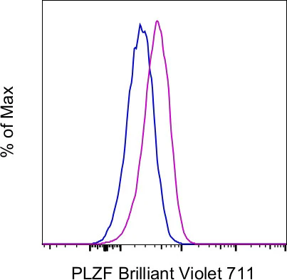
Applications Tested: This Mags.21F7 antibody has been tested by intracellular staining followed by flow cytometric analysis of mouse thymocytes using the Foxp3/Transcription Factor Staining Buffer Set (Product # 00-5523-00) and protocol. Please refer to "Staining Intracellular Antigens for Flow Cytometry, Protocol B: One step protocol for intracellular (nuclear) proteins" located at Flow Protocols. This may be used at less than or equal to 1.0 µg per test. A test is defined as the amount (µg) of antibody that will stain a cell sample in a final volume of 100 µL. Cell number should be determined empirically but can range from 10^5 to 10^8 cells/test. It is recommended that the antibody be carefully titrated for optimal performance in the assay of interest.
Brilliant Violet™ 711 (BV711) is a tandem dye that emits at 713 nm and is intended for use on cytometers equipped with a violet (405 nm) laser. Please make sure that your instrument is capable of detecting this fluorochrome.
When using two or more Super Bright, Brilliant Violet™, Brilliant Ultra Violet™, or other polymer dye-conjugated antibodies in a staining panel, it is recommended to use Super Bright Complete Staining Buffer (Product # SB-4401-42) or Brilliant Stain Buffer™ (Product # 00-4409-75) to minimize any non-specific polymer interactions. Please refer to the datasheet for Super Bright Staining Buffer or Brilliant Stain Buffer for more information.
Light sensitivity: This tandem dye is sensitive to photo-induced oxidation. Please protect this vial and stained samples from light.
Fixation: Samples can be stored in IC Fixation Buffer (Product # 00-8222-49) (100 µL of cell sample + 100 µL of IC Fixation Buffer) or 1-step Fix/Lyse Solution (Product # 00-5333-54) for up to 3 days in the dark at 4°C with minimal impact on brightness and FRET efficiency/compensation. Some generalizations regarding fluorophore performance after fixation can be made, but clone-specific performance should be determined empirically.
Our internal testing suggests that Brilliant Violet™ 711 (BV711) is not compatible with methanol-based fixation.
Excitation: 407 nm; Emission: 713 nm; Laser: Violet Laser.
BRILLIANT VIOLET™ is a trademark or registered trademark of Becton, Dickinson and Company or its affiliates, and is used under license. Powered by Sirigen™.
For Research Use Only. Not for use in diagnostic procedures. Not for resale without express authorization.
Original: $194.00
-70%$194.00
$58.20PLZF Monoclonal Antibody (Mags.21F7), Brilliant Violet 711, eBioscience
PRODUCT DETAILS
Host: Mouse
Isotype: IgG1, kappa
Clonality: Monoclonal
Clone: Mags.21F7
Format: Brilliant Violet 711
Reactivity: Hu, Ms
Application: Flow Cytometry
Tested Dilution: 1.0 µg/test
Concentration: 0.2 mg/mL
Storage: 4°C, store in dark, DO NOT FREEZE!
Formulation: PBS with BSA and 0.09% sodium azide; pH 7.2
Purification: Affinity chromatography
Data Sheet: TDS
Specific Information
Description: This Mags.21F7 monoclonal antibody reacts with human and mouse promyelocytic leukemia zinc finger (PLZF), a member of the BTB-POZ family of transcription factors. Expression of this transcriptional repressor in immune cells differs between mice and humans. In mice, PLZF is highly expressed in immature CD1d-resricted NKT2 and NKT1 cells, and a subset of gamma delta (Vg1.1+Vd6.3+) T cells. Studies have also demonstrated expression of PLZF in non-invariant CD1d-restricted T cells, as well as non-CD1d-restricted innate T cells. In humans, PLZF is expressed in NK cells, gamma delta T cells, as well as CD4 and CD8+ T cells. PLZF is also expressed in MR1-specific mucosal-associated invariant T cells, as well as in MHC Class II-restricted T cells that develop via a thymocyte-thymocyte interaction in humans. PLZF exists as a homodimer or in complex with PLZP, and has been shown to be involved in the development of NKT cells, NK cell function, cellular quiescence, and growth suppression. Finally, PLZF has been shown to inhibit gene expression induced by retinoic acid receptor.
Applications Reported: This Mags.21F7 antibody has been reported for use in intracellular staining followed by flow cytometric analysis.
Applications Tested: This Mags.21F7 antibody has been tested by intracellular staining followed by flow cytometric analysis of mouse thymocytes using the Foxp3/Transcription Factor Staining Buffer Set (Product # 00-5523-00) and protocol. Please refer to "Staining Intracellular Antigens for Flow Cytometry, Protocol B: One step protocol for intracellular (nuclear) proteins" located at Flow Protocols. This may be used at less than or equal to 1.0 µg per test. A test is defined as the amount (µg) of antibody that will stain a cell sample in a final volume of 100 µL. Cell number should be determined empirically but can range from 10^5 to 10^8 cells/test. It is recommended that the antibody be carefully titrated for optimal performance in the assay of interest.
Brilliant Violet™ 711 (BV711) is a tandem dye that emits at 713 nm and is intended for use on cytometers equipped with a violet (405 nm) laser. Please make sure that your instrument is capable of detecting this fluorochrome.
When using two or more Super Bright, Brilliant Violet™, Brilliant Ultra Violet™, or other polymer dye-conjugated antibodies in a staining panel, it is recommended to use Super Bright Complete Staining Buffer (Product # SB-4401-42) or Brilliant Stain Buffer™ (Product # 00-4409-75) to minimize any non-specific polymer interactions. Please refer to the datasheet for Super Bright Staining Buffer or Brilliant Stain Buffer for more information.
Light sensitivity: This tandem dye is sensitive to photo-induced oxidation. Please protect this vial and stained samples from light.
Fixation: Samples can be stored in IC Fixation Buffer (Product # 00-8222-49) (100 µL of cell sample + 100 µL of IC Fixation Buffer) or 1-step Fix/Lyse Solution (Product # 00-5333-54) for up to 3 days in the dark at 4°C with minimal impact on brightness and FRET efficiency/compensation. Some generalizations regarding fluorophore performance after fixation can be made, but clone-specific performance should be determined empirically.
Our internal testing suggests that Brilliant Violet™ 711 (BV711) is not compatible with methanol-based fixation.
Excitation: 407 nm; Emission: 713 nm; Laser: Violet Laser.
BRILLIANT VIOLET™ is a trademark or registered trademark of Becton, Dickinson and Company or its affiliates, and is used under license. Powered by Sirigen™.
For Research Use Only. Not for use in diagnostic procedures. Not for resale without express authorization.
Product Information
Product Information
Shipping & Returns
Shipping & Returns
Description
PRODUCT DETAILS
Host: Mouse
Isotype: IgG1, kappa
Clonality: Monoclonal
Clone: Mags.21F7
Format: Brilliant Violet 711
Reactivity: Hu, Ms
Application: Flow Cytometry
Tested Dilution: 1.0 µg/test
Concentration: 0.2 mg/mL
Storage: 4°C, store in dark, DO NOT FREEZE!
Formulation: PBS with BSA and 0.09% sodium azide; pH 7.2
Purification: Affinity chromatography
Data Sheet: TDS
Specific Information
Description: This Mags.21F7 monoclonal antibody reacts with human and mouse promyelocytic leukemia zinc finger (PLZF), a member of the BTB-POZ family of transcription factors. Expression of this transcriptional repressor in immune cells differs between mice and humans. In mice, PLZF is highly expressed in immature CD1d-resricted NKT2 and NKT1 cells, and a subset of gamma delta (Vg1.1+Vd6.3+) T cells. Studies have also demonstrated expression of PLZF in non-invariant CD1d-restricted T cells, as well as non-CD1d-restricted innate T cells. In humans, PLZF is expressed in NK cells, gamma delta T cells, as well as CD4 and CD8+ T cells. PLZF is also expressed in MR1-specific mucosal-associated invariant T cells, as well as in MHC Class II-restricted T cells that develop via a thymocyte-thymocyte interaction in humans. PLZF exists as a homodimer or in complex with PLZP, and has been shown to be involved in the development of NKT cells, NK cell function, cellular quiescence, and growth suppression. Finally, PLZF has been shown to inhibit gene expression induced by retinoic acid receptor.
Applications Reported: This Mags.21F7 antibody has been reported for use in intracellular staining followed by flow cytometric analysis.
Applications Tested: This Mags.21F7 antibody has been tested by intracellular staining followed by flow cytometric analysis of mouse thymocytes using the Foxp3/Transcription Factor Staining Buffer Set (Product # 00-5523-00) and protocol. Please refer to "Staining Intracellular Antigens for Flow Cytometry, Protocol B: One step protocol for intracellular (nuclear) proteins" located at Flow Protocols. This may be used at less than or equal to 1.0 µg per test. A test is defined as the amount (µg) of antibody that will stain a cell sample in a final volume of 100 µL. Cell number should be determined empirically but can range from 10^5 to 10^8 cells/test. It is recommended that the antibody be carefully titrated for optimal performance in the assay of interest.
Brilliant Violet™ 711 (BV711) is a tandem dye that emits at 713 nm and is intended for use on cytometers equipped with a violet (405 nm) laser. Please make sure that your instrument is capable of detecting this fluorochrome.
When using two or more Super Bright, Brilliant Violet™, Brilliant Ultra Violet™, or other polymer dye-conjugated antibodies in a staining panel, it is recommended to use Super Bright Complete Staining Buffer (Product # SB-4401-42) or Brilliant Stain Buffer™ (Product # 00-4409-75) to minimize any non-specific polymer interactions. Please refer to the datasheet for Super Bright Staining Buffer or Brilliant Stain Buffer for more information.
Light sensitivity: This tandem dye is sensitive to photo-induced oxidation. Please protect this vial and stained samples from light.
Fixation: Samples can be stored in IC Fixation Buffer (Product # 00-8222-49) (100 µL of cell sample + 100 µL of IC Fixation Buffer) or 1-step Fix/Lyse Solution (Product # 00-5333-54) for up to 3 days in the dark at 4°C with minimal impact on brightness and FRET efficiency/compensation. Some generalizations regarding fluorophore performance after fixation can be made, but clone-specific performance should be determined empirically.
Our internal testing suggests that Brilliant Violet™ 711 (BV711) is not compatible with methanol-based fixation.
Excitation: 407 nm; Emission: 713 nm; Laser: Violet Laser.
BRILLIANT VIOLET™ is a trademark or registered trademark of Becton, Dickinson and Company or its affiliates, and is used under license. Powered by Sirigen™.
For Research Use Only. Not for use in diagnostic procedures. Not for resale without express authorization.


