phospho-Tyrosine Monoclonal Antibody (pY20), eFluor 660, eBioscience
PRODUCT DETAILS
Host: Mouse
Isotype: IgG2b, kappa
Clonality: Monoclonal
Clone: pY20
Format: eFluor 660
Reactivity: Chem
Application: Flow Cytometry
Tested Dilution: 5 µL (0.06 µg)/test
Concentration: 5 μL/Test
Storage: 4°C, store in dark, DO NOT FREEZE!
Formulation: PBS with BSA and 0.09% sodium azide; pH 7.2
Purification: Affinity chromatography
Data Sheet: TDS
Specific Information
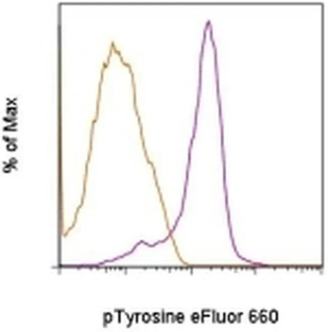
Description: The pY20 monoclonal antibody recognizes phosphorylated tyrosine residues (p-Tyr) on proteins. Numerous intracellular signaling cascades are propagated via phosphorylation of specific tyrosine on signaling proteins. The detection of p-Tyr residues is valuable for the characterization and purification of phosphorylated proteins and the biochemical pathways that they are involved in.
Applications Reported:This pY20 antibody has been reported for use in intracellular staining followed by flow cytometric analysis.
Applications Tested: This pY20 antibody has been pre-titrated and tested by intracellular staining followed by flow cytometric analysis of normal human peripheral blood cells. This can be used at 5 µL (0.06 µg) per test. A test is defined as the amount (µg) of antibody that will stain a cell sample in a final volume of 100 µL. Cell number should be determined empirically but can range from 10^5 to 10^8 cells/test.
Staining Protocol: All protocols work well for this monoclonal antibody. Use of Protocol A: Two-step protocol: intracellular (cytoplasmic) proteins allows for the greatest flexibility for detection of surface and intracellular (cytoplasmic) proteins. Use of Protocol B: One-step protocol: intracellular (nuclear) proteins is recommended for staining of transcription factors in conjunction with surface and phosphorylated intracellular (cytoplasmic) protein(s). Protocol C: Two-step protocol: Fixation/Methanol allows for the greatest discrimination of phospho-specific signaling between unstimulated and stimulated samples, but with limitations on the ability to stain specific surface proteins (refer to "Clone Performance Following Fixation/Permeabilization" located in the BestProtocols Section under the Resources tab online). All Protocols can be found in the Flow Cytometry Protocols: "Staining Intracellular Antigens for Flow Cytometry Protocol" located in the BestProtocols® Section under the Resources tab online.
Excitation: 633-647 nm; Emission: 668 nm; Laser: Red Laser.
Filtration: 0.2 µm post-manufacturing filtered.
For Research Use Only. Not for use in diagnostic procedures. Not for resale without express authorization.
phospho-Tyrosine Monoclonal Antibody (pY20), eFluor 660, eBioscience
PRODUCT DETAILS
Host: Mouse
Isotype: IgG2b, kappa
Clonality: Monoclonal
Clone: pY20
Format: eFluor 660
Reactivity: Chem
Application: Flow Cytometry
Tested Dilution: 5 µL (0.06 µg)/test
Concentration: 5 μL/Test
Storage: 4°C, store in dark, DO NOT FREEZE!
Formulation: PBS with BSA and 0.09% sodium azide; pH 7.2
Purification: Affinity chromatography
Data Sheet: TDS
Specific Information
Description: The pY20 monoclonal antibody recognizes phosphorylated tyrosine residues (p-Tyr) on proteins. Numerous intracellular signaling cascades are propagated via phosphorylation of specific tyrosine on signaling proteins. The detection of p-Tyr residues is valuable for the characterization and purification of phosphorylated proteins and the biochemical pathways that they are involved in.
Applications Reported:This pY20 antibody has been reported for use in intracellular staining followed by flow cytometric analysis.
Applications Tested: This pY20 antibody has been pre-titrated and tested by intracellular staining followed by flow cytometric analysis of normal human peripheral blood cells. This can be used at 5 µL (0.06 µg) per test. A test is defined as the amount (µg) of antibody that will stain a cell sample in a final volume of 100 µL. Cell number should be determined empirically but can range from 10^5 to 10^8 cells/test.
Staining Protocol: All protocols work well for this monoclonal antibody. Use of Protocol A: Two-step protocol: intracellular (cytoplasmic) proteins allows for the greatest flexibility for detection of surface and intracellular (cytoplasmic) proteins. Use of Protocol B: One-step protocol: intracellular (nuclear) proteins is recommended for staining of transcription factors in conjunction with surface and phosphorylated intracellular (cytoplasmic) protein(s). Protocol C: Two-step protocol: Fixation/Methanol allows for the greatest discrimination of phospho-specific signaling between unstimulated and stimulated samples, but with limitations on the ability to stain specific surface proteins (refer to "Clone Performance Following Fixation/Permeabilization" located in the BestProtocols Section under the Resources tab online). All Protocols can be found in the Flow Cytometry Protocols: "Staining Intracellular Antigens for Flow Cytometry Protocol" located in the BestProtocols® Section under the Resources tab online.
Excitation: 633-647 nm; Emission: 668 nm; Laser: Red Laser.
Filtration: 0.2 µm post-manufacturing filtered.
For Research Use Only. Not for use in diagnostic procedures. Not for resale without express authorization.
Product Information
Product Information
Shipping & Returns
Shipping & Returns
Description
PRODUCT DETAILS
Host: Mouse
Isotype: IgG2b, kappa
Clonality: Monoclonal
Clone: pY20
Format: eFluor 660
Reactivity: Chem
Application: Flow Cytometry
Tested Dilution: 5 µL (0.06 µg)/test
Concentration: 5 μL/Test
Storage: 4°C, store in dark, DO NOT FREEZE!
Formulation: PBS with BSA and 0.09% sodium azide; pH 7.2
Purification: Affinity chromatography
Data Sheet: TDS
Specific Information
Description: The pY20 monoclonal antibody recognizes phosphorylated tyrosine residues (p-Tyr) on proteins. Numerous intracellular signaling cascades are propagated via phosphorylation of specific tyrosine on signaling proteins. The detection of p-Tyr residues is valuable for the characterization and purification of phosphorylated proteins and the biochemical pathways that they are involved in.
Applications Reported:This pY20 antibody has been reported for use in intracellular staining followed by flow cytometric analysis.
Applications Tested: This pY20 antibody has been pre-titrated and tested by intracellular staining followed by flow cytometric analysis of normal human peripheral blood cells. This can be used at 5 µL (0.06 µg) per test. A test is defined as the amount (µg) of antibody that will stain a cell sample in a final volume of 100 µL. Cell number should be determined empirically but can range from 10^5 to 10^8 cells/test.
Staining Protocol: All protocols work well for this monoclonal antibody. Use of Protocol A: Two-step protocol: intracellular (cytoplasmic) proteins allows for the greatest flexibility for detection of surface and intracellular (cytoplasmic) proteins. Use of Protocol B: One-step protocol: intracellular (nuclear) proteins is recommended for staining of transcription factors in conjunction with surface and phosphorylated intracellular (cytoplasmic) protein(s). Protocol C: Two-step protocol: Fixation/Methanol allows for the greatest discrimination of phospho-specific signaling between unstimulated and stimulated samples, but with limitations on the ability to stain specific surface proteins (refer to "Clone Performance Following Fixation/Permeabilization" located in the BestProtocols Section under the Resources tab online). All Protocols can be found in the Flow Cytometry Protocols: "Staining Intracellular Antigens for Flow Cytometry Protocol" located in the BestProtocols® Section under the Resources tab online.
Excitation: 633-647 nm; Emission: 668 nm; Laser: Red Laser.
Filtration: 0.2 µm post-manufacturing filtered.
For Research Use Only. Not for use in diagnostic procedures. Not for resale without express authorization.


