Phospho-Syk (Tyr348) Monoclonal Antibody (moch1ct), APC, eBioscience
PRODUCT DETAILS
Host: Mouse
Isotype: IgG1, kappa
Clonality: Monoclonal
Clone: moch1ct
Format: APC
Reactivity: Hu, Ms
Application: Flow Cytometry
Tested Dilution: 5 µL (0.06 µg)/test
Concentration: 5 μL/Test
Storage: 4°C, store in dark, DO NOT FREEZE!
Formulation: PBS with BSA and 0.09% sodium azide; pH 7.2
Purification: Affinity chromatography
Data Sheet: TDS
Specific Information
Description: This moch1ct monoclonal antibody recognizes human and mouse spleen tyrosine kinase (also known as SYK) when phosphorylated on tyrosine 348 (Y348). SYK is the founding member of the SYK family of kinases that also includes ZAP-70 (zeta-associated protein of 70 kD) and is expressed in hematopoietic cells, including B lymphocytes, immature (CD4, CD8 double-negative and double-positive) thymocytes, and myeloid cells, epithelial cell lines, and normal breast tissue. SYK is critical for B cell receptor (BCR) signaling and B cell development. Autophosphorylation of at Y348 is necessary for SYK to become fully catalytically active and creates a docking site for the SH2 domains of Vav, Grb2, p85 subunit of PI3 kinase, and PLC gamma.
Specificity of this moch1ct clone was confirmed by ELISA, flow cytometry, and western blotting.
Applications Reported:This moch1ct antibody has been reported for use in intracellular staining followed by flow cytometric analysis.
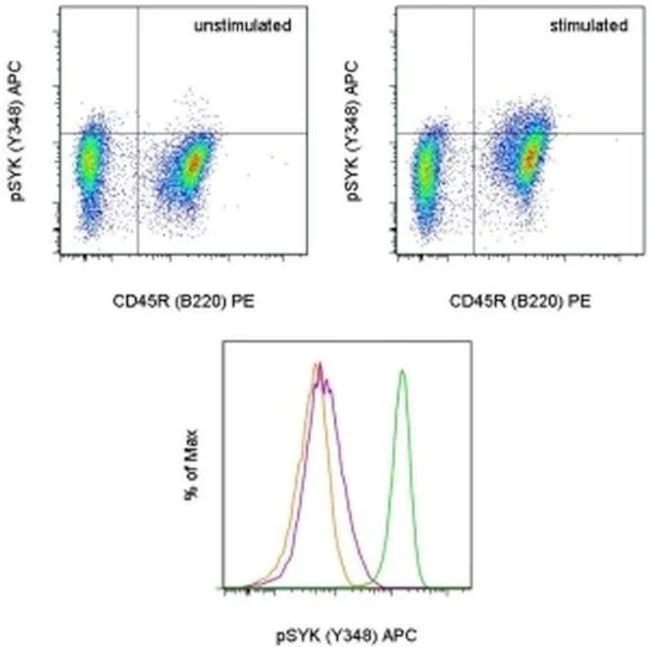
Applications Tested: This moch1ct antibody has been pre-titrated and tested by intracellular staining followed by flow cytometric analysis of stimulated mouse splenocytes. This can be used at 5 µL (0.06 µg) per test. A test is defined as the amount (µg) of antibody that will stain a cell sample in a final volume of 100 µL. Cell number should be determined empirically but can range from 10^5 to 10^8 cells/test.
Staining Protocol: All protocols work well for this monoclonal antibody. Use of Protocol A: Two-step protocol: intracellular (cytoplasmic) proteins allows for the greatest flexibility for detection of surface and intracellular (cytoplasmic) proteins. Use of Protocol B: One-step protocol: intracellular (nuclear) proteins is recommended for staining of transcription factors in conjunction with surface and phosphorylated intracellular (cytoplasmic) proteins. Protocol C: Two-step protocol: Fixation/Methanol allows for the greatest discrimination of phospho-specific signaling between unstimulated and stimulated samples, but with limitations on the ability to stain specific surface proteins (refer to "Clone Performance Following Fixation/Permeabilization" located in the BestProtocols Section under the Resources tab online). All Protocols can be found in the Flow Cytometry Protocols: "Staining Intracellular Antigens for Flow Cytometry Protocol" located in the BestProtocols® Section under the Resources tab online.
Excitation: 633-647 nm; Emission: 660 nm; Laser: Red Laser.
Filtration: 0.2 µm post-manufacturing filtered.
For Research Use Only. Not for use in diagnostic procedures. Not for resale without express authorization.
Original: $446.00
-70%$446.00
$133.80Phospho-Syk (Tyr348) Monoclonal Antibody (moch1ct), APC, eBioscience
PRODUCT DETAILS
Host: Mouse
Isotype: IgG1, kappa
Clonality: Monoclonal
Clone: moch1ct
Format: APC
Reactivity: Hu, Ms
Application: Flow Cytometry
Tested Dilution: 5 µL (0.06 µg)/test
Concentration: 5 μL/Test
Storage: 4°C, store in dark, DO NOT FREEZE!
Formulation: PBS with BSA and 0.09% sodium azide; pH 7.2
Purification: Affinity chromatography
Data Sheet: TDS
Specific Information
Description: This moch1ct monoclonal antibody recognizes human and mouse spleen tyrosine kinase (also known as SYK) when phosphorylated on tyrosine 348 (Y348). SYK is the founding member of the SYK family of kinases that also includes ZAP-70 (zeta-associated protein of 70 kD) and is expressed in hematopoietic cells, including B lymphocytes, immature (CD4, CD8 double-negative and double-positive) thymocytes, and myeloid cells, epithelial cell lines, and normal breast tissue. SYK is critical for B cell receptor (BCR) signaling and B cell development. Autophosphorylation of at Y348 is necessary for SYK to become fully catalytically active and creates a docking site for the SH2 domains of Vav, Grb2, p85 subunit of PI3 kinase, and PLC gamma.
Specificity of this moch1ct clone was confirmed by ELISA, flow cytometry, and western blotting.
Applications Reported:This moch1ct antibody has been reported for use in intracellular staining followed by flow cytometric analysis.
Applications Tested: This moch1ct antibody has been pre-titrated and tested by intracellular staining followed by flow cytometric analysis of stimulated mouse splenocytes. This can be used at 5 µL (0.06 µg) per test. A test is defined as the amount (µg) of antibody that will stain a cell sample in a final volume of 100 µL. Cell number should be determined empirically but can range from 10^5 to 10^8 cells/test.
Staining Protocol: All protocols work well for this monoclonal antibody. Use of Protocol A: Two-step protocol: intracellular (cytoplasmic) proteins allows for the greatest flexibility for detection of surface and intracellular (cytoplasmic) proteins. Use of Protocol B: One-step protocol: intracellular (nuclear) proteins is recommended for staining of transcription factors in conjunction with surface and phosphorylated intracellular (cytoplasmic) proteins. Protocol C: Two-step protocol: Fixation/Methanol allows for the greatest discrimination of phospho-specific signaling between unstimulated and stimulated samples, but with limitations on the ability to stain specific surface proteins (refer to "Clone Performance Following Fixation/Permeabilization" located in the BestProtocols Section under the Resources tab online). All Protocols can be found in the Flow Cytometry Protocols: "Staining Intracellular Antigens for Flow Cytometry Protocol" located in the BestProtocols® Section under the Resources tab online.
Excitation: 633-647 nm; Emission: 660 nm; Laser: Red Laser.
Filtration: 0.2 µm post-manufacturing filtered.
For Research Use Only. Not for use in diagnostic procedures. Not for resale without express authorization.
Product Information
Product Information
Shipping & Returns
Shipping & Returns
Description
PRODUCT DETAILS
Host: Mouse
Isotype: IgG1, kappa
Clonality: Monoclonal
Clone: moch1ct
Format: APC
Reactivity: Hu, Ms
Application: Flow Cytometry
Tested Dilution: 5 µL (0.06 µg)/test
Concentration: 5 μL/Test
Storage: 4°C, store in dark, DO NOT FREEZE!
Formulation: PBS with BSA and 0.09% sodium azide; pH 7.2
Purification: Affinity chromatography
Data Sheet: TDS
Specific Information
Description: This moch1ct monoclonal antibody recognizes human and mouse spleen tyrosine kinase (also known as SYK) when phosphorylated on tyrosine 348 (Y348). SYK is the founding member of the SYK family of kinases that also includes ZAP-70 (zeta-associated protein of 70 kD) and is expressed in hematopoietic cells, including B lymphocytes, immature (CD4, CD8 double-negative and double-positive) thymocytes, and myeloid cells, epithelial cell lines, and normal breast tissue. SYK is critical for B cell receptor (BCR) signaling and B cell development. Autophosphorylation of at Y348 is necessary for SYK to become fully catalytically active and creates a docking site for the SH2 domains of Vav, Grb2, p85 subunit of PI3 kinase, and PLC gamma.
Specificity of this moch1ct clone was confirmed by ELISA, flow cytometry, and western blotting.
Applications Reported:This moch1ct antibody has been reported for use in intracellular staining followed by flow cytometric analysis.
Applications Tested: This moch1ct antibody has been pre-titrated and tested by intracellular staining followed by flow cytometric analysis of stimulated mouse splenocytes. This can be used at 5 µL (0.06 µg) per test. A test is defined as the amount (µg) of antibody that will stain a cell sample in a final volume of 100 µL. Cell number should be determined empirically but can range from 10^5 to 10^8 cells/test.
Staining Protocol: All protocols work well for this monoclonal antibody. Use of Protocol A: Two-step protocol: intracellular (cytoplasmic) proteins allows for the greatest flexibility for detection of surface and intracellular (cytoplasmic) proteins. Use of Protocol B: One-step protocol: intracellular (nuclear) proteins is recommended for staining of transcription factors in conjunction with surface and phosphorylated intracellular (cytoplasmic) proteins. Protocol C: Two-step protocol: Fixation/Methanol allows for the greatest discrimination of phospho-specific signaling between unstimulated and stimulated samples, but with limitations on the ability to stain specific surface proteins (refer to "Clone Performance Following Fixation/Permeabilization" located in the BestProtocols Section under the Resources tab online). All Protocols can be found in the Flow Cytometry Protocols: "Staining Intracellular Antigens for Flow Cytometry Protocol" located in the BestProtocols® Section under the Resources tab online.
Excitation: 633-647 nm; Emission: 660 nm; Laser: Red Laser.
Filtration: 0.2 µm post-manufacturing filtered.
For Research Use Only. Not for use in diagnostic procedures. Not for resale without express authorization.


