Phospho-STAT4 (Tyr693) Monoclonal Antibody (4LURPIE), APC, eBioscience
PRODUCT DETAILS
Host: Mouse
Isotype: IgG1, kappa
Clonality: Monoclonal
Clone: 4LURPIE
Format: APC
Reactivity: Hu, Ms
Application: Flow Cytometry
Tested Dilution: 5 µL (0.25 µg)/test
Concentration: 5 μL/Test
Storage: 4°C, store in dark, DO NOT FREEZE!
Formulation: PBS with BSA and 0.09% sodium azide; pH 7.2
Purification: Affinity chromatography
Data Sheet: TDS
Specific Information
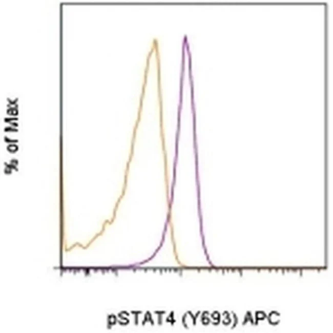
Description: This 4LURPIE monoclonal antibody recognizes human and mouse signal transducer and activator of transcription 4 (STAT4) when phosphorylated on tyrosine 693 (Y693). STAT proteins are activated by ligand binding to cytokine receptors that associate with Janus kinase (JAK) family members. Following their phosphorylation by JAK proteins, STAT proteins translocate to the nucleus where they bind to DNA and regulate transcription of specific genes in a cell type- and cytokine-specific manner. STAT4 is activated by IL-12 or type 1 IFN (IFN alpha, IFN beta). STAT4 is required for optimal Th1 differentiation in vivo. Interestingly, although IFN alpha can lead to phosphorylation of STAT4 in mouse T cells, the phosphorylation is much weaker than that induced by IL-12 and is transient. Thus, IFN alpha phosphorylation of STAT4 does not contribute to STAT4-mediated Th1 development or IFN gamma production by Th1 cells in mice. In contrast, IFN alpha does promote Th1 development and function of human CD4+ T cells.
Specificity of this 4LURPIE clone was determined by ELISA and flow cytometry.
Applications Reported:This 4LURPIE antibody has been reported for use in intracellular staining followed by flow cytometric analysis.
Applications Tested: This 4LURPIE antibody has been pre-titrated and tested by intracellular staining followed by flow cytometric analysis of stimulated normal human peripheral blood cells. This can be used at 5 µL (0.25 µg) per test. A test is defined as the amount (µg) of antibody that will stain a cell sample in a final volume of 100 µL. Cell number should be determined empirically but can range from 10^5 to 10^8 cells/test.
Staining Protocol: We recommend using Protocol C: Two-step protocol: Fixation/Methanol. Protocol A: Two-step protocol: intracellular (cytoplasmic) proteins and Protocol B: One-step protocol: intracellular (nuclear) proteins cannot be used. All Protocols can be found in the Flow Cytometry Protocols: "Staining Intracellular Antigens for Flow Cytometry Protocol" located in the BestProtocols® Section under the Resources tab online.
Excitation: 633-647 nm; Emission: 660 nm; Laser: Red Laser.
Filtration: 0.2 µm post-manufacturing filtered.
For Research Use Only. Not for use in diagnostic procedures. Not for resale without express authorization.
Original: $434.00
-70%$434.00
$130.20Phospho-STAT4 (Tyr693) Monoclonal Antibody (4LURPIE), APC, eBioscience
PRODUCT DETAILS
Host: Mouse
Isotype: IgG1, kappa
Clonality: Monoclonal
Clone: 4LURPIE
Format: APC
Reactivity: Hu, Ms
Application: Flow Cytometry
Tested Dilution: 5 µL (0.25 µg)/test
Concentration: 5 μL/Test
Storage: 4°C, store in dark, DO NOT FREEZE!
Formulation: PBS with BSA and 0.09% sodium azide; pH 7.2
Purification: Affinity chromatography
Data Sheet: TDS
Specific Information
Description: This 4LURPIE monoclonal antibody recognizes human and mouse signal transducer and activator of transcription 4 (STAT4) when phosphorylated on tyrosine 693 (Y693). STAT proteins are activated by ligand binding to cytokine receptors that associate with Janus kinase (JAK) family members. Following their phosphorylation by JAK proteins, STAT proteins translocate to the nucleus where they bind to DNA and regulate transcription of specific genes in a cell type- and cytokine-specific manner. STAT4 is activated by IL-12 or type 1 IFN (IFN alpha, IFN beta). STAT4 is required for optimal Th1 differentiation in vivo. Interestingly, although IFN alpha can lead to phosphorylation of STAT4 in mouse T cells, the phosphorylation is much weaker than that induced by IL-12 and is transient. Thus, IFN alpha phosphorylation of STAT4 does not contribute to STAT4-mediated Th1 development or IFN gamma production by Th1 cells in mice. In contrast, IFN alpha does promote Th1 development and function of human CD4+ T cells.
Specificity of this 4LURPIE clone was determined by ELISA and flow cytometry.
Applications Reported:This 4LURPIE antibody has been reported for use in intracellular staining followed by flow cytometric analysis.
Applications Tested: This 4LURPIE antibody has been pre-titrated and tested by intracellular staining followed by flow cytometric analysis of stimulated normal human peripheral blood cells. This can be used at 5 µL (0.25 µg) per test. A test is defined as the amount (µg) of antibody that will stain a cell sample in a final volume of 100 µL. Cell number should be determined empirically but can range from 10^5 to 10^8 cells/test.
Staining Protocol: We recommend using Protocol C: Two-step protocol: Fixation/Methanol. Protocol A: Two-step protocol: intracellular (cytoplasmic) proteins and Protocol B: One-step protocol: intracellular (nuclear) proteins cannot be used. All Protocols can be found in the Flow Cytometry Protocols: "Staining Intracellular Antigens for Flow Cytometry Protocol" located in the BestProtocols® Section under the Resources tab online.
Excitation: 633-647 nm; Emission: 660 nm; Laser: Red Laser.
Filtration: 0.2 µm post-manufacturing filtered.
For Research Use Only. Not for use in diagnostic procedures. Not for resale without express authorization.
Product Information
Product Information
Shipping & Returns
Shipping & Returns
Description
PRODUCT DETAILS
Host: Mouse
Isotype: IgG1, kappa
Clonality: Monoclonal
Clone: 4LURPIE
Format: APC
Reactivity: Hu, Ms
Application: Flow Cytometry
Tested Dilution: 5 µL (0.25 µg)/test
Concentration: 5 μL/Test
Storage: 4°C, store in dark, DO NOT FREEZE!
Formulation: PBS with BSA and 0.09% sodium azide; pH 7.2
Purification: Affinity chromatography
Data Sheet: TDS
Specific Information
Description: This 4LURPIE monoclonal antibody recognizes human and mouse signal transducer and activator of transcription 4 (STAT4) when phosphorylated on tyrosine 693 (Y693). STAT proteins are activated by ligand binding to cytokine receptors that associate with Janus kinase (JAK) family members. Following their phosphorylation by JAK proteins, STAT proteins translocate to the nucleus where they bind to DNA and regulate transcription of specific genes in a cell type- and cytokine-specific manner. STAT4 is activated by IL-12 or type 1 IFN (IFN alpha, IFN beta). STAT4 is required for optimal Th1 differentiation in vivo. Interestingly, although IFN alpha can lead to phosphorylation of STAT4 in mouse T cells, the phosphorylation is much weaker than that induced by IL-12 and is transient. Thus, IFN alpha phosphorylation of STAT4 does not contribute to STAT4-mediated Th1 development or IFN gamma production by Th1 cells in mice. In contrast, IFN alpha does promote Th1 development and function of human CD4+ T cells.
Specificity of this 4LURPIE clone was determined by ELISA and flow cytometry.
Applications Reported:This 4LURPIE antibody has been reported for use in intracellular staining followed by flow cytometric analysis.
Applications Tested: This 4LURPIE antibody has been pre-titrated and tested by intracellular staining followed by flow cytometric analysis of stimulated normal human peripheral blood cells. This can be used at 5 µL (0.25 µg) per test. A test is defined as the amount (µg) of antibody that will stain a cell sample in a final volume of 100 µL. Cell number should be determined empirically but can range from 10^5 to 10^8 cells/test.
Staining Protocol: We recommend using Protocol C: Two-step protocol: Fixation/Methanol. Protocol A: Two-step protocol: intracellular (cytoplasmic) proteins and Protocol B: One-step protocol: intracellular (nuclear) proteins cannot be used. All Protocols can be found in the Flow Cytometry Protocols: "Staining Intracellular Antigens for Flow Cytometry Protocol" located in the BestProtocols® Section under the Resources tab online.
Excitation: 633-647 nm; Emission: 660 nm; Laser: Red Laser.
Filtration: 0.2 µm post-manufacturing filtered.
For Research Use Only. Not for use in diagnostic procedures. Not for resale without express authorization.


