Phospho-SLP-76 (Tyr128) Monoclonal Antibody (HNDZ55), APC, eBioscience
PRODUCT DETAILS
Host: Mouse
Isotype: IgG2b, kappa
Clonality: Monoclonal
Clone: HNDZ55
Format: APC
Reactivity: Hu
Application: Flow Cytometry
Tested Dilution: 5 µL (0.5 µg)/test
Concentration: 5 μL/Test
Storage: 4°C, store in dark, DO NOT FREEZE!
Formulation: PBS with BSA and 0.09% sodium azide; pH 7.2
Purification: Affinity chromatography
Data Sheet: TDS
Specific Information
Description: This HNDZ55 monoclonal antibody recognizes human SH2 domain-containing leukocyte protein of 76 kD (SLP-76) when phosphorylated on tyrosine 128 (Y128). SLP-76 is phosphorylated by ZAP-70 downstream of the T cell receptor (TCR). SLP-76 contains an acidic region that includes a PEST domain, several tyrosine residues that are phosphorylated following TCR ligation, a proline-rich domain, and an SH2 domain. Numerous proteins associate with SLP-76 including Vav1, GADS, and PLC gamma 1, supporting the notion that SLP-76 functions as a critical adaptor protein downstream of the TCR. SLP-76-deficient T cell lines or mice deficient in SLP-76 have shown that SLP-76 plays a positive role in promoting T cell development and activation as well as mast cell and platelet function.
This antibody recognizes only human SLP-76 when phosphorylated on Y128. It is not recommended for staining of mouse cells.
Applications Reported:This HNDZ55 antibody has been reported for use in intracellular staining followed by flow cytometric analysis.
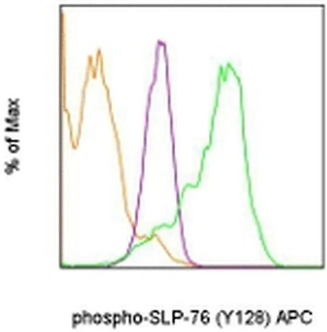
Applications Tested: This HNDZ55 antibody has been pre-titrated and tested by intracellular staining followed by flow cytometric analysis of stimulated normal human peripheral blood cells. This can be used at 5 µL (0.5 µg) per test. A test is defined as the amount (µg) of antibody that will stain a cell sample in a final volume of 100 µL. Cell number should be determined empirically but can range from 10^5 to 10^8 cells/test.
Staining Protocol: Protocol A and Protocol C are recommended for this monoclonal antibody. Use of Protocol A: Two-step protocol: intracellular (cytoplasmic) proteins allows for the greatest flexibility for detection of surface and intracellular (cytoplasmic) proteins. Protocol C: Two-step protocol: Fixation/Methanol allows for the greatest discrimination of phospho-specific signaling between unstimulated and stimulated samples, but with limitations on the ability to stain specific surface proteins (refer to "Clone Performance Following Fixation/Permeabilization" located in the BestProtocols Section under the Resources tab online). All Protocols can be found in the Flow Cytometry Protocols: "Staining Intracellular Antigens for Flow Cytometry Protocol" located in the BestProtocols® Section under the Resources tab online.
Excitation: 633-647 nm; Emission: 660 nm; Laser: Red Laser.
Filtration: 0.2 µm post-manufacturing filtered.
For Research Use Only. Not for use in diagnostic procedures. Not for resale without express authorization.
Original: $438.00
-70%$438.00
$131.40Phospho-SLP-76 (Tyr128) Monoclonal Antibody (HNDZ55), APC, eBioscience
PRODUCT DETAILS
Host: Mouse
Isotype: IgG2b, kappa
Clonality: Monoclonal
Clone: HNDZ55
Format: APC
Reactivity: Hu
Application: Flow Cytometry
Tested Dilution: 5 µL (0.5 µg)/test
Concentration: 5 μL/Test
Storage: 4°C, store in dark, DO NOT FREEZE!
Formulation: PBS with BSA and 0.09% sodium azide; pH 7.2
Purification: Affinity chromatography
Data Sheet: TDS
Specific Information
Description: This HNDZ55 monoclonal antibody recognizes human SH2 domain-containing leukocyte protein of 76 kD (SLP-76) when phosphorylated on tyrosine 128 (Y128). SLP-76 is phosphorylated by ZAP-70 downstream of the T cell receptor (TCR). SLP-76 contains an acidic region that includes a PEST domain, several tyrosine residues that are phosphorylated following TCR ligation, a proline-rich domain, and an SH2 domain. Numerous proteins associate with SLP-76 including Vav1, GADS, and PLC gamma 1, supporting the notion that SLP-76 functions as a critical adaptor protein downstream of the TCR. SLP-76-deficient T cell lines or mice deficient in SLP-76 have shown that SLP-76 plays a positive role in promoting T cell development and activation as well as mast cell and platelet function.
This antibody recognizes only human SLP-76 when phosphorylated on Y128. It is not recommended for staining of mouse cells.
Applications Reported:This HNDZ55 antibody has been reported for use in intracellular staining followed by flow cytometric analysis.
Applications Tested: This HNDZ55 antibody has been pre-titrated and tested by intracellular staining followed by flow cytometric analysis of stimulated normal human peripheral blood cells. This can be used at 5 µL (0.5 µg) per test. A test is defined as the amount (µg) of antibody that will stain a cell sample in a final volume of 100 µL. Cell number should be determined empirically but can range from 10^5 to 10^8 cells/test.
Staining Protocol: Protocol A and Protocol C are recommended for this monoclonal antibody. Use of Protocol A: Two-step protocol: intracellular (cytoplasmic) proteins allows for the greatest flexibility for detection of surface and intracellular (cytoplasmic) proteins. Protocol C: Two-step protocol: Fixation/Methanol allows for the greatest discrimination of phospho-specific signaling between unstimulated and stimulated samples, but with limitations on the ability to stain specific surface proteins (refer to "Clone Performance Following Fixation/Permeabilization" located in the BestProtocols Section under the Resources tab online). All Protocols can be found in the Flow Cytometry Protocols: "Staining Intracellular Antigens for Flow Cytometry Protocol" located in the BestProtocols® Section under the Resources tab online.
Excitation: 633-647 nm; Emission: 660 nm; Laser: Red Laser.
Filtration: 0.2 µm post-manufacturing filtered.
For Research Use Only. Not for use in diagnostic procedures. Not for resale without express authorization.
Product Information
Product Information
Shipping & Returns
Shipping & Returns
Description
PRODUCT DETAILS
Host: Mouse
Isotype: IgG2b, kappa
Clonality: Monoclonal
Clone: HNDZ55
Format: APC
Reactivity: Hu
Application: Flow Cytometry
Tested Dilution: 5 µL (0.5 µg)/test
Concentration: 5 μL/Test
Storage: 4°C, store in dark, DO NOT FREEZE!
Formulation: PBS with BSA and 0.09% sodium azide; pH 7.2
Purification: Affinity chromatography
Data Sheet: TDS
Specific Information
Description: This HNDZ55 monoclonal antibody recognizes human SH2 domain-containing leukocyte protein of 76 kD (SLP-76) when phosphorylated on tyrosine 128 (Y128). SLP-76 is phosphorylated by ZAP-70 downstream of the T cell receptor (TCR). SLP-76 contains an acidic region that includes a PEST domain, several tyrosine residues that are phosphorylated following TCR ligation, a proline-rich domain, and an SH2 domain. Numerous proteins associate with SLP-76 including Vav1, GADS, and PLC gamma 1, supporting the notion that SLP-76 functions as a critical adaptor protein downstream of the TCR. SLP-76-deficient T cell lines or mice deficient in SLP-76 have shown that SLP-76 plays a positive role in promoting T cell development and activation as well as mast cell and platelet function.
This antibody recognizes only human SLP-76 when phosphorylated on Y128. It is not recommended for staining of mouse cells.
Applications Reported:This HNDZ55 antibody has been reported for use in intracellular staining followed by flow cytometric analysis.
Applications Tested: This HNDZ55 antibody has been pre-titrated and tested by intracellular staining followed by flow cytometric analysis of stimulated normal human peripheral blood cells. This can be used at 5 µL (0.5 µg) per test. A test is defined as the amount (µg) of antibody that will stain a cell sample in a final volume of 100 µL. Cell number should be determined empirically but can range from 10^5 to 10^8 cells/test.
Staining Protocol: Protocol A and Protocol C are recommended for this monoclonal antibody. Use of Protocol A: Two-step protocol: intracellular (cytoplasmic) proteins allows for the greatest flexibility for detection of surface and intracellular (cytoplasmic) proteins. Protocol C: Two-step protocol: Fixation/Methanol allows for the greatest discrimination of phospho-specific signaling between unstimulated and stimulated samples, but with limitations on the ability to stain specific surface proteins (refer to "Clone Performance Following Fixation/Permeabilization" located in the BestProtocols Section under the Resources tab online). All Protocols can be found in the Flow Cytometry Protocols: "Staining Intracellular Antigens for Flow Cytometry Protocol" located in the BestProtocols® Section under the Resources tab online.
Excitation: 633-647 nm; Emission: 660 nm; Laser: Red Laser.
Filtration: 0.2 µm post-manufacturing filtered.
For Research Use Only. Not for use in diagnostic procedures. Not for resale without express authorization.


