Phospho-NFkB p65 (Ser529) Monoclonal Antibody (B33B4WP), PE, eBioscience
PRODUCT DETAILS
Host: Mouse
Isotype: IgG2a, kappa
Clonality: Monoclonal
Clone: B33B4WP
Format: PE
Reactivity: Hu
Application: Flow Cytometry
Tested Dilution: 5 µL (0.06 µg)/test
Concentration: 5 μL/Test
Storage: 4°C, store in dark, DO NOT FREEZE!
Formulation: PBS with BSA and 0.09% sodium azide; pH 7.2
Purification: Affinity chromatography
Data Sheet: TDS
Specific Information
Description: The B33B4WP monoclonal antibody recognizes human NF kappa B (NFkB) p65 subunit when serine 529 is phosphorylated. NFkB, also known as nuclear factor kappa-light chain enhancer of activated B cells, is a ubiquitous transcription factor that regulates the transcription of many genes involved in cell proliferation, apoptosis, development, immunity and cancer. Functional NFkB is a homo- or hetero-dimer composed of 5 members of the NFkB family: p65 (RelA), c-Rel, RelB, p50 (NFkB1, p105 precursor protein), and p52 (NFkB2, p100 precursor protein). The activity of the complex is negatively regulated by binding to IkB inhibitors that sequester NFkB into the cytoplasm, inhibiting its transcriptional activity. NFkB-activating agents like tumor necrosis factor (TNF) alpha, interleukin-1 beta, lipopolysaccharide, camptothecin, and phorbol ester (PMA) induce the phosphorylation and degradation of IkB, leading to the translocation of NFkB to the nucleus where it binds to kB motifs and regulates gene expression. The activity of p65-containing NFkB complexes is positively regulated by phosphorylation of the p65 subunit at serine 529 and/or serine 536.
The B33B4WP monoclonal antibody recognizes a single band at 65 kDa in reduced SDS lysates from TNF alpha-stimulated HeLa cells.
Applications Reported:This B33B4WP antibody has been reported for use in intracellular staining followed by flow cytometric analysis.
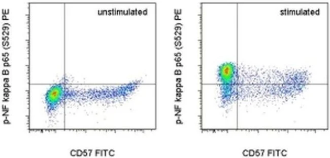
Applications Tested: This B33B4WP antibody has been pre-titrated and tested by intracellular staining followed by flow cytometric analysis of stimulated normal human peripheral blood cells. This can be used at 5 µL (0.06 µg) per test. A test is defined as the amount (µg) of antibody that will stain a cell sample in a final volume of 100 µL. Cell number should be determined empirically but can range from 10^5 to 10^8 cells/test.
Protocols: We recommend Protocol A: Two-step protocol: intracellular (cytoplasmic) proteins. Alternatively, Protocol C: Two-step protocol: Fixation/Methanol can also be used. Protocol B: One-step protocol: intracellular (nuclear) proteins cannot be used. All Protocols can be found in the "Staining intracellular Antigens for Flow Cytometry Protocol" located in the BestProtocols® Section under the Resources tab online.
Excitation: 488-561 nm; Emission: 578 nm; Laser: Blue Laser, Green Laser, Yellow-Green Laser.
Filtration: 0.2 µm post-manufacturing filtered.
For Research Use Only. Not for use in diagnostic procedures. Not for resale without express authorization.
Phospho-NFkB p65 (Ser529) Monoclonal Antibody (B33B4WP), PE, eBioscience
PRODUCT DETAILS
Host: Mouse
Isotype: IgG2a, kappa
Clonality: Monoclonal
Clone: B33B4WP
Format: PE
Reactivity: Hu
Application: Flow Cytometry
Tested Dilution: 5 µL (0.06 µg)/test
Concentration: 5 μL/Test
Storage: 4°C, store in dark, DO NOT FREEZE!
Formulation: PBS with BSA and 0.09% sodium azide; pH 7.2
Purification: Affinity chromatography
Data Sheet: TDS
Specific Information
Description: The B33B4WP monoclonal antibody recognizes human NF kappa B (NFkB) p65 subunit when serine 529 is phosphorylated. NFkB, also known as nuclear factor kappa-light chain enhancer of activated B cells, is a ubiquitous transcription factor that regulates the transcription of many genes involved in cell proliferation, apoptosis, development, immunity and cancer. Functional NFkB is a homo- or hetero-dimer composed of 5 members of the NFkB family: p65 (RelA), c-Rel, RelB, p50 (NFkB1, p105 precursor protein), and p52 (NFkB2, p100 precursor protein). The activity of the complex is negatively regulated by binding to IkB inhibitors that sequester NFkB into the cytoplasm, inhibiting its transcriptional activity. NFkB-activating agents like tumor necrosis factor (TNF) alpha, interleukin-1 beta, lipopolysaccharide, camptothecin, and phorbol ester (PMA) induce the phosphorylation and degradation of IkB, leading to the translocation of NFkB to the nucleus where it binds to kB motifs and regulates gene expression. The activity of p65-containing NFkB complexes is positively regulated by phosphorylation of the p65 subunit at serine 529 and/or serine 536.
The B33B4WP monoclonal antibody recognizes a single band at 65 kDa in reduced SDS lysates from TNF alpha-stimulated HeLa cells.
Applications Reported:This B33B4WP antibody has been reported for use in intracellular staining followed by flow cytometric analysis.
Applications Tested: This B33B4WP antibody has been pre-titrated and tested by intracellular staining followed by flow cytometric analysis of stimulated normal human peripheral blood cells. This can be used at 5 µL (0.06 µg) per test. A test is defined as the amount (µg) of antibody that will stain a cell sample in a final volume of 100 µL. Cell number should be determined empirically but can range from 10^5 to 10^8 cells/test.
Protocols: We recommend Protocol A: Two-step protocol: intracellular (cytoplasmic) proteins. Alternatively, Protocol C: Two-step protocol: Fixation/Methanol can also be used. Protocol B: One-step protocol: intracellular (nuclear) proteins cannot be used. All Protocols can be found in the "Staining intracellular Antigens for Flow Cytometry Protocol" located in the BestProtocols® Section under the Resources tab online.
Excitation: 488-561 nm; Emission: 578 nm; Laser: Blue Laser, Green Laser, Yellow-Green Laser.
Filtration: 0.2 µm post-manufacturing filtered.
For Research Use Only. Not for use in diagnostic procedures. Not for resale without express authorization.
Product Information
Product Information
Shipping & Returns
Shipping & Returns
Description
PRODUCT DETAILS
Host: Mouse
Isotype: IgG2a, kappa
Clonality: Monoclonal
Clone: B33B4WP
Format: PE
Reactivity: Hu
Application: Flow Cytometry
Tested Dilution: 5 µL (0.06 µg)/test
Concentration: 5 μL/Test
Storage: 4°C, store in dark, DO NOT FREEZE!
Formulation: PBS with BSA and 0.09% sodium azide; pH 7.2
Purification: Affinity chromatography
Data Sheet: TDS
Specific Information
Description: The B33B4WP monoclonal antibody recognizes human NF kappa B (NFkB) p65 subunit when serine 529 is phosphorylated. NFkB, also known as nuclear factor kappa-light chain enhancer of activated B cells, is a ubiquitous transcription factor that regulates the transcription of many genes involved in cell proliferation, apoptosis, development, immunity and cancer. Functional NFkB is a homo- or hetero-dimer composed of 5 members of the NFkB family: p65 (RelA), c-Rel, RelB, p50 (NFkB1, p105 precursor protein), and p52 (NFkB2, p100 precursor protein). The activity of the complex is negatively regulated by binding to IkB inhibitors that sequester NFkB into the cytoplasm, inhibiting its transcriptional activity. NFkB-activating agents like tumor necrosis factor (TNF) alpha, interleukin-1 beta, lipopolysaccharide, camptothecin, and phorbol ester (PMA) induce the phosphorylation and degradation of IkB, leading to the translocation of NFkB to the nucleus where it binds to kB motifs and regulates gene expression. The activity of p65-containing NFkB complexes is positively regulated by phosphorylation of the p65 subunit at serine 529 and/or serine 536.
The B33B4WP monoclonal antibody recognizes a single band at 65 kDa in reduced SDS lysates from TNF alpha-stimulated HeLa cells.
Applications Reported:This B33B4WP antibody has been reported for use in intracellular staining followed by flow cytometric analysis.
Applications Tested: This B33B4WP antibody has been pre-titrated and tested by intracellular staining followed by flow cytometric analysis of stimulated normal human peripheral blood cells. This can be used at 5 µL (0.06 µg) per test. A test is defined as the amount (µg) of antibody that will stain a cell sample in a final volume of 100 µL. Cell number should be determined empirically but can range from 10^5 to 10^8 cells/test.
Protocols: We recommend Protocol A: Two-step protocol: intracellular (cytoplasmic) proteins. Alternatively, Protocol C: Two-step protocol: Fixation/Methanol can also be used. Protocol B: One-step protocol: intracellular (nuclear) proteins cannot be used. All Protocols can be found in the "Staining intracellular Antigens for Flow Cytometry Protocol" located in the BestProtocols® Section under the Resources tab online.
Excitation: 488-561 nm; Emission: 578 nm; Laser: Blue Laser, Green Laser, Yellow-Green Laser.
Filtration: 0.2 µm post-manufacturing filtered.
For Research Use Only. Not for use in diagnostic procedures. Not for resale without express authorization.


