Phospho-LCK (Tyr505) Monoclonal Antibody (SRRCHA), PerCP-eFluor 710, eBioscience
PRODUCT DETAILS
Host: Mouse
Isotype: IgG1, kappa
Clonality: Monoclonal
Clone: SRRCHA
Format: PerCP-eFluor 710
Reactivity: Hu, Ms
Application: Flow Cytometry
Tested Dilution: 5 µL (0.06 µg)/test
Concentration: 5 μL/Test
Storage: 4°C, store in dark, DO NOT FREEZE!
Formulation: PBS with BSA and 0.09% sodium azide; pH 7.2
Purification: Affinity chromatography
Data Sheet: TDS
Specific Information
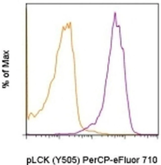
Description: This SRRCHA monoclonal antibody recognizes human and mouse LCK when phosphorylated on tyrosine 505 (Y505). Y505 is a negative-regulatory site that is phosphorylated by the kinase CSK. LCK is expressed in T cells where it associates with the cytoplasmic tails of the CD4 and CD8 co-receptors on T helper cells and cytotoxic T cells, respectively. When the TCR is engaged, LCK acts to phosphorylate the intracellular chains of CD3 and zeta chains of the TCR complex, allowing the cytoplasmic tyrosine kinase, ZAP-70, to bind to them. LCK then phosphorylates and activates ZAP-70, which then phosphorylates the adaptor protein, LAT, that serves as a docking site for a number of other proteins, including SLP-76, Grb2, and phospholipase C gamma1. Mice lacking expression of LCK show defects in T cell development and function.
Applications Reported: This SRRCHA antibody has been reported for use in intracellular staining followed by flow cytometric analysis.
Applications Tested: This SRRCHA antibody has been pre-titrated and tested by intracellular staining followed by flow cytometric analysis of activated human peripheral blood cells. This can be used at 5 µL (0.06 µg) per test. A test is defined as the amount (µg) of antibody that will stain a cell sample in a final volume of 100 µL. Cell number should be determined empirically but can range from 10^5 to 10^8 cells/test.
PerCP-eFluor® 710 emits at 710 nm and is excited with the blue laser (488 nm); it can be used in place of PerCP-Cyanine5.5. We recommend using a 710/50 bandpass filter, however, the 695/40 bandpass filter is an acceptable alternative. Please make sure that your instrument is capable of detecting this fluorochrome.
Light sensitivity: This tandem dye is sensitive to photo-induced oxidation. Please protect this vial and stained samples from light.
Fixation: Samples can be stored in IC Fixation Buffer (Product # 00-8222) (100 µL of cell sample + 100 µL of IC Fixation Buffer) or 1-step Fix/Lyse Solution (Product # 00-5333) for up to 3 days in the dark at 4°C with minimal impact on brightness and FRET efficiency/compensation. Some generalizations regarding fluorophore performance after fixation can be made, but clone specific performance should be determined empirically.
Staining Protocol: All protocols work well for this monoclonal antibody. Use of Protocol A: Two-step protocol: intracellular (cytoplasmic) proteins allows for the greatest flexibility for detection of surface and intracellular (cytoplasmic) proteins. Use of Protocol B: One-step protocol: intracellular (nuclear) proteins is recommended for staining of transcription factors in conjunction with surface and phosphorylated intracellular (cytoplasmic) proteins. Protocol C: Two-step protocol: Fixation/Methanol allows for the greatest discrimination of phospho-specific signaling between unstimulated and stimulated samples, but with limitations on the ability to stain specific surface proteins (refer to "Clone Performance Following Fixation/Permeabilization" located in the BestProtocols Section under the Resources tab online). All Protocols can be found in the Flow Cytometry Protocols: "Staining Intracellular Antigens for Flow Cytometry Protocol" located in the BestProtocols® Section under the Resources tab online.
Excitation: 488 nm; Emission: 710 nm; Laser: Blue Laser.
Filtration: 0.2 µm post-manufacturing filtered.
For Research Use Only. Not for use in diagnostic procedures. Not for resale without express authorization.
Phospho-LCK (Tyr505) Monoclonal Antibody (SRRCHA), PerCP-eFluor 710, eBioscience
PRODUCT DETAILS
Host: Mouse
Isotype: IgG1, kappa
Clonality: Monoclonal
Clone: SRRCHA
Format: PerCP-eFluor 710
Reactivity: Hu, Ms
Application: Flow Cytometry
Tested Dilution: 5 µL (0.06 µg)/test
Concentration: 5 μL/Test
Storage: 4°C, store in dark, DO NOT FREEZE!
Formulation: PBS with BSA and 0.09% sodium azide; pH 7.2
Purification: Affinity chromatography
Data Sheet: TDS
Specific Information
Description: This SRRCHA monoclonal antibody recognizes human and mouse LCK when phosphorylated on tyrosine 505 (Y505). Y505 is a negative-regulatory site that is phosphorylated by the kinase CSK. LCK is expressed in T cells where it associates with the cytoplasmic tails of the CD4 and CD8 co-receptors on T helper cells and cytotoxic T cells, respectively. When the TCR is engaged, LCK acts to phosphorylate the intracellular chains of CD3 and zeta chains of the TCR complex, allowing the cytoplasmic tyrosine kinase, ZAP-70, to bind to them. LCK then phosphorylates and activates ZAP-70, which then phosphorylates the adaptor protein, LAT, that serves as a docking site for a number of other proteins, including SLP-76, Grb2, and phospholipase C gamma1. Mice lacking expression of LCK show defects in T cell development and function.
Applications Reported: This SRRCHA antibody has been reported for use in intracellular staining followed by flow cytometric analysis.
Applications Tested: This SRRCHA antibody has been pre-titrated and tested by intracellular staining followed by flow cytometric analysis of activated human peripheral blood cells. This can be used at 5 µL (0.06 µg) per test. A test is defined as the amount (µg) of antibody that will stain a cell sample in a final volume of 100 µL. Cell number should be determined empirically but can range from 10^5 to 10^8 cells/test.
PerCP-eFluor® 710 emits at 710 nm and is excited with the blue laser (488 nm); it can be used in place of PerCP-Cyanine5.5. We recommend using a 710/50 bandpass filter, however, the 695/40 bandpass filter is an acceptable alternative. Please make sure that your instrument is capable of detecting this fluorochrome.
Light sensitivity: This tandem dye is sensitive to photo-induced oxidation. Please protect this vial and stained samples from light.
Fixation: Samples can be stored in IC Fixation Buffer (Product # 00-8222) (100 µL of cell sample + 100 µL of IC Fixation Buffer) or 1-step Fix/Lyse Solution (Product # 00-5333) for up to 3 days in the dark at 4°C with minimal impact on brightness and FRET efficiency/compensation. Some generalizations regarding fluorophore performance after fixation can be made, but clone specific performance should be determined empirically.
Staining Protocol: All protocols work well for this monoclonal antibody. Use of Protocol A: Two-step protocol: intracellular (cytoplasmic) proteins allows for the greatest flexibility for detection of surface and intracellular (cytoplasmic) proteins. Use of Protocol B: One-step protocol: intracellular (nuclear) proteins is recommended for staining of transcription factors in conjunction with surface and phosphorylated intracellular (cytoplasmic) proteins. Protocol C: Two-step protocol: Fixation/Methanol allows for the greatest discrimination of phospho-specific signaling between unstimulated and stimulated samples, but with limitations on the ability to stain specific surface proteins (refer to "Clone Performance Following Fixation/Permeabilization" located in the BestProtocols Section under the Resources tab online). All Protocols can be found in the Flow Cytometry Protocols: "Staining Intracellular Antigens for Flow Cytometry Protocol" located in the BestProtocols® Section under the Resources tab online.
Excitation: 488 nm; Emission: 710 nm; Laser: Blue Laser.
Filtration: 0.2 µm post-manufacturing filtered.
For Research Use Only. Not for use in diagnostic procedures. Not for resale without express authorization.
Product Information
Product Information
Shipping & Returns
Shipping & Returns
Description
PRODUCT DETAILS
Host: Mouse
Isotype: IgG1, kappa
Clonality: Monoclonal
Clone: SRRCHA
Format: PerCP-eFluor 710
Reactivity: Hu, Ms
Application: Flow Cytometry
Tested Dilution: 5 µL (0.06 µg)/test
Concentration: 5 μL/Test
Storage: 4°C, store in dark, DO NOT FREEZE!
Formulation: PBS with BSA and 0.09% sodium azide; pH 7.2
Purification: Affinity chromatography
Data Sheet: TDS
Specific Information
Description: This SRRCHA monoclonal antibody recognizes human and mouse LCK when phosphorylated on tyrosine 505 (Y505). Y505 is a negative-regulatory site that is phosphorylated by the kinase CSK. LCK is expressed in T cells where it associates with the cytoplasmic tails of the CD4 and CD8 co-receptors on T helper cells and cytotoxic T cells, respectively. When the TCR is engaged, LCK acts to phosphorylate the intracellular chains of CD3 and zeta chains of the TCR complex, allowing the cytoplasmic tyrosine kinase, ZAP-70, to bind to them. LCK then phosphorylates and activates ZAP-70, which then phosphorylates the adaptor protein, LAT, that serves as a docking site for a number of other proteins, including SLP-76, Grb2, and phospholipase C gamma1. Mice lacking expression of LCK show defects in T cell development and function.
Applications Reported: This SRRCHA antibody has been reported for use in intracellular staining followed by flow cytometric analysis.
Applications Tested: This SRRCHA antibody has been pre-titrated and tested by intracellular staining followed by flow cytometric analysis of activated human peripheral blood cells. This can be used at 5 µL (0.06 µg) per test. A test is defined as the amount (µg) of antibody that will stain a cell sample in a final volume of 100 µL. Cell number should be determined empirically but can range from 10^5 to 10^8 cells/test.
PerCP-eFluor® 710 emits at 710 nm and is excited with the blue laser (488 nm); it can be used in place of PerCP-Cyanine5.5. We recommend using a 710/50 bandpass filter, however, the 695/40 bandpass filter is an acceptable alternative. Please make sure that your instrument is capable of detecting this fluorochrome.
Light sensitivity: This tandem dye is sensitive to photo-induced oxidation. Please protect this vial and stained samples from light.
Fixation: Samples can be stored in IC Fixation Buffer (Product # 00-8222) (100 µL of cell sample + 100 µL of IC Fixation Buffer) or 1-step Fix/Lyse Solution (Product # 00-5333) for up to 3 days in the dark at 4°C with minimal impact on brightness and FRET efficiency/compensation. Some generalizations regarding fluorophore performance after fixation can be made, but clone specific performance should be determined empirically.
Staining Protocol: All protocols work well for this monoclonal antibody. Use of Protocol A: Two-step protocol: intracellular (cytoplasmic) proteins allows for the greatest flexibility for detection of surface and intracellular (cytoplasmic) proteins. Use of Protocol B: One-step protocol: intracellular (nuclear) proteins is recommended for staining of transcription factors in conjunction with surface and phosphorylated intracellular (cytoplasmic) proteins. Protocol C: Two-step protocol: Fixation/Methanol allows for the greatest discrimination of phospho-specific signaling between unstimulated and stimulated samples, but with limitations on the ability to stain specific surface proteins (refer to "Clone Performance Following Fixation/Permeabilization" located in the BestProtocols Section under the Resources tab online). All Protocols can be found in the Flow Cytometry Protocols: "Staining Intracellular Antigens for Flow Cytometry Protocol" located in the BestProtocols® Section under the Resources tab online.
Excitation: 488 nm; Emission: 710 nm; Laser: Blue Laser.
Filtration: 0.2 µm post-manufacturing filtered.
For Research Use Only. Not for use in diagnostic procedures. Not for resale without express authorization.


