Phospho-LCK (Tyr505) Monoclonal Antibody (SRRCHA), PE, eBioscience
PRODUCT DETAILS
Host: Mouse
Isotype: IgG1, kappa
Clonality: Monoclonal
Clone: SRRCHA
Format: PE
Reactivity: Hu, Ms
Application: Flow Cytometry
Tested Dilution: 5 µL (0.25 µg)/test
Concentration: 5 μL/Test
Storage: 4°C, store in dark, DO NOT FREEZE!
Formulation: PBS with BSA and 0.09% sodium azide; pH 7.2
Purification: Affinity chromatography
Data Sheet: TDS
Specific Information
Description: This SRRCHA monoclonal antibody recognizes human and mouse LCK when phosphorylated on tyrosine 505 (Y505). Y505 is a negative-regulatory site that is phosphorylated by the kinase CSK. LCK is expressed in T cells where it associates with the cytoplasmic tails of the CD4 and CD8 co-receptors on T helper cells and cytotoxic T cells, respectively. When the TCR is engaged, LCK acts to phosphorylate the intracellular chains of CD3 and zeta chains of the TCR complex, allowing the cytoplasmic tyrosine kinase, ZAP-70, to bind to them. LCK then phosphorylates and activates ZAP-70, which then phosphorylates the adaptor protein, LAT, that serves as a docking site for a number of other proteins, including SLP-76, Grb2, and phospholipase C gamma1. Mice lacking expression of LCK show defects in T cell development and function.
Applications Reported: This SRRCHA antibody has been reported for use in intracellular staining followed by flow cytometric analysis.
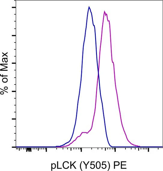
Applications Tested: This SRRCHA antibody has been pre-diluted and tested by intracellular staining followed by flow cytometric analysis of Jurkat cells using the Intracellular Fixation & Permeabilization Buffer Set (Product # 88-8824-00) and protocol. Please refer to "Staining Intracellular Antigens for Flow Cytometry, Protocol A: Two step protocol for intracellular (cytoplasmic) proteins" located in the BestProtocols® Section under the Resources tab online. This may be used at 5 µL (0.25 µg) per test. A test is defined as the amount (µg) of antibody that will stain a cell sample in a final volume of 100 µL. Cell number should be determined empirically but can range from 10^5 to 10^8 cells/test.
Excitation: 488-561 nm; Emission: 578 nm; Laser: Blue Laser, Green Laser, Yellow-Green Laser.
For Research Use Only. Not for use in diagnostic procedures. Not for resale without express authorization.
Phospho-LCK (Tyr505) Monoclonal Antibody (SRRCHA), PE, eBioscience
PRODUCT DETAILS
Host: Mouse
Isotype: IgG1, kappa
Clonality: Monoclonal
Clone: SRRCHA
Format: PE
Reactivity: Hu, Ms
Application: Flow Cytometry
Tested Dilution: 5 µL (0.25 µg)/test
Concentration: 5 μL/Test
Storage: 4°C, store in dark, DO NOT FREEZE!
Formulation: PBS with BSA and 0.09% sodium azide; pH 7.2
Purification: Affinity chromatography
Data Sheet: TDS
Specific Information
Description: This SRRCHA monoclonal antibody recognizes human and mouse LCK when phosphorylated on tyrosine 505 (Y505). Y505 is a negative-regulatory site that is phosphorylated by the kinase CSK. LCK is expressed in T cells where it associates with the cytoplasmic tails of the CD4 and CD8 co-receptors on T helper cells and cytotoxic T cells, respectively. When the TCR is engaged, LCK acts to phosphorylate the intracellular chains of CD3 and zeta chains of the TCR complex, allowing the cytoplasmic tyrosine kinase, ZAP-70, to bind to them. LCK then phosphorylates and activates ZAP-70, which then phosphorylates the adaptor protein, LAT, that serves as a docking site for a number of other proteins, including SLP-76, Grb2, and phospholipase C gamma1. Mice lacking expression of LCK show defects in T cell development and function.
Applications Reported: This SRRCHA antibody has been reported for use in intracellular staining followed by flow cytometric analysis.
Applications Tested: This SRRCHA antibody has been pre-diluted and tested by intracellular staining followed by flow cytometric analysis of Jurkat cells using the Intracellular Fixation & Permeabilization Buffer Set (Product # 88-8824-00) and protocol. Please refer to "Staining Intracellular Antigens for Flow Cytometry, Protocol A: Two step protocol for intracellular (cytoplasmic) proteins" located in the BestProtocols® Section under the Resources tab online. This may be used at 5 µL (0.25 µg) per test. A test is defined as the amount (µg) of antibody that will stain a cell sample in a final volume of 100 µL. Cell number should be determined empirically but can range from 10^5 to 10^8 cells/test.
Excitation: 488-561 nm; Emission: 578 nm; Laser: Blue Laser, Green Laser, Yellow-Green Laser.
For Research Use Only. Not for use in diagnostic procedures. Not for resale without express authorization.
Product Information
Product Information
Shipping & Returns
Shipping & Returns
Description
PRODUCT DETAILS
Host: Mouse
Isotype: IgG1, kappa
Clonality: Monoclonal
Clone: SRRCHA
Format: PE
Reactivity: Hu, Ms
Application: Flow Cytometry
Tested Dilution: 5 µL (0.25 µg)/test
Concentration: 5 μL/Test
Storage: 4°C, store in dark, DO NOT FREEZE!
Formulation: PBS with BSA and 0.09% sodium azide; pH 7.2
Purification: Affinity chromatography
Data Sheet: TDS
Specific Information
Description: This SRRCHA monoclonal antibody recognizes human and mouse LCK when phosphorylated on tyrosine 505 (Y505). Y505 is a negative-regulatory site that is phosphorylated by the kinase CSK. LCK is expressed in T cells where it associates with the cytoplasmic tails of the CD4 and CD8 co-receptors on T helper cells and cytotoxic T cells, respectively. When the TCR is engaged, LCK acts to phosphorylate the intracellular chains of CD3 and zeta chains of the TCR complex, allowing the cytoplasmic tyrosine kinase, ZAP-70, to bind to them. LCK then phosphorylates and activates ZAP-70, which then phosphorylates the adaptor protein, LAT, that serves as a docking site for a number of other proteins, including SLP-76, Grb2, and phospholipase C gamma1. Mice lacking expression of LCK show defects in T cell development and function.
Applications Reported: This SRRCHA antibody has been reported for use in intracellular staining followed by flow cytometric analysis.
Applications Tested: This SRRCHA antibody has been pre-diluted and tested by intracellular staining followed by flow cytometric analysis of Jurkat cells using the Intracellular Fixation & Permeabilization Buffer Set (Product # 88-8824-00) and protocol. Please refer to "Staining Intracellular Antigens for Flow Cytometry, Protocol A: Two step protocol for intracellular (cytoplasmic) proteins" located in the BestProtocols® Section under the Resources tab online. This may be used at 5 µL (0.25 µg) per test. A test is defined as the amount (µg) of antibody that will stain a cell sample in a final volume of 100 µL. Cell number should be determined empirically but can range from 10^5 to 10^8 cells/test.
Excitation: 488-561 nm; Emission: 578 nm; Laser: Blue Laser, Green Laser, Yellow-Green Laser.
For Research Use Only. Not for use in diagnostic procedures. Not for resale without express authorization.


