Phospho-IkB alpha (Ser32, Ser36) Monoclonal Antibody (RILYB3R), PE, eBioscience
PRODUCT DETAILS
Host: Mouse
Isotype: IgG2b, kappa
Clonality: Monoclonal
Clone: RILYB3R
Format: PE
Reactivity: Hu, Ms
Application: Flow Cytometry
Tested Dilution: 5 µL (0.125 µg)/test
Concentration: 5 μL/Test
Storage: 4°C, store in dark, DO NOT FREEZE!
Formulation: PBS with BSA and 0.09% sodium azide; pH 7.2
Purification: Affinity chromatography
Data Sheet: TDS
Specific Information
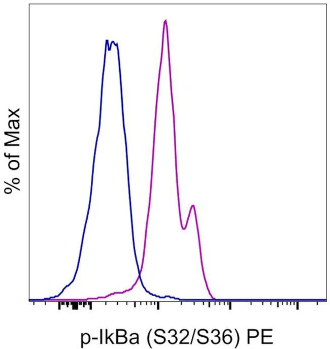
Description: This RILYB3R monoclonal antibody recognizes human and mouse nuclear factor of kappa light polypeptide gene enhancer in B cells inhibitor, alpha (I kappa B alpha) when phosphorylated on serines 32 and 36 (S32/S36). I kappa B alpha is one member of a family of cellular proteins that functions to inhibit classical/canonical NF-kappa B signaling by masking the nuclear localization signals (NLS) of NF-kappa B proteins and keeping them sequestered in an inactive state in the cytoplasm. Classical/canonical NF-kappa B signaling is initiated in response to myriad stimuli including engagement of T cell and B cell receptors, growth factors, and inflammatory stimuli (reactive oxygen species, TNF alpha, IL-1) and results in the activation of the I kappa B kinase (IKK) complex that includes IKK alpha, IKK beta, and NEMO. IKK phosphorylates I kappa B alpha resulting in its ubiquitination, degradation, and subsequent translocation of NF-kappa B transcription factor proteins into the nucleus.
Applications Reported: This RILYB3R antibody has been reported for use in intracellular staining followed by flow cytometric analysis.
Applications Tested: This RILYB3R antibody has been pre-titrated and tested by intracellular staining followed by flow cytometric analysis of normal human peripheral blood cells. This can be used at 5 µL (0.125 µg) per test. A test is defined as the amount (µg) of antibody that will stain a cell sample in a final volume of 100 µL. Cell number should be determined empirically but can range from 10^5 to 10^8 cells/test. It is recommended that the antibody be carefully titrated for optimal performance in the assay of interest.
Staining Protocol: Protocol A and Protocol C are recommended for this monoclonal antibody. Use of Protocol A: Two-step protocol: intracellular (cytoplasmic) proteins allows for the greatest flexibility for detection of surface and intracellular (cytoplasmic) proteins. Protocol C: Two-step protocol: Fixation/Methanol allows for the greatest discrimination of phospho-specific signaling between unstimulated and stimulated samples, but with limitations on the ability to stain specific surface proteins (refer to "Clone Performance Following Fixation/Permeabilization" located in the BestProtocols Section under the Resources tab online). All Protocols can be found in the Flow Cytometry Protocols: "Staining Intracellular Antigens for Flow Cytometry Protocol" located in the BestProtocols® Section under the Resources tab online.
Emission: 578 nm; Laser: Blue Laser, Green Laser, Yellow-Green Laser.
Filtration: 0.2 µm post-manufacturing filtered.
For Research Use Only. Not for use in diagnostic procedures. Not for resale without express authorization.
Phospho-IkB alpha (Ser32, Ser36) Monoclonal Antibody (RILYB3R), PE, eBioscience
PRODUCT DETAILS
Host: Mouse
Isotype: IgG2b, kappa
Clonality: Monoclonal
Clone: RILYB3R
Format: PE
Reactivity: Hu, Ms
Application: Flow Cytometry
Tested Dilution: 5 µL (0.125 µg)/test
Concentration: 5 μL/Test
Storage: 4°C, store in dark, DO NOT FREEZE!
Formulation: PBS with BSA and 0.09% sodium azide; pH 7.2
Purification: Affinity chromatography
Data Sheet: TDS
Specific Information
Description: This RILYB3R monoclonal antibody recognizes human and mouse nuclear factor of kappa light polypeptide gene enhancer in B cells inhibitor, alpha (I kappa B alpha) when phosphorylated on serines 32 and 36 (S32/S36). I kappa B alpha is one member of a family of cellular proteins that functions to inhibit classical/canonical NF-kappa B signaling by masking the nuclear localization signals (NLS) of NF-kappa B proteins and keeping them sequestered in an inactive state in the cytoplasm. Classical/canonical NF-kappa B signaling is initiated in response to myriad stimuli including engagement of T cell and B cell receptors, growth factors, and inflammatory stimuli (reactive oxygen species, TNF alpha, IL-1) and results in the activation of the I kappa B kinase (IKK) complex that includes IKK alpha, IKK beta, and NEMO. IKK phosphorylates I kappa B alpha resulting in its ubiquitination, degradation, and subsequent translocation of NF-kappa B transcription factor proteins into the nucleus.
Applications Reported: This RILYB3R antibody has been reported for use in intracellular staining followed by flow cytometric analysis.
Applications Tested: This RILYB3R antibody has been pre-titrated and tested by intracellular staining followed by flow cytometric analysis of normal human peripheral blood cells. This can be used at 5 µL (0.125 µg) per test. A test is defined as the amount (µg) of antibody that will stain a cell sample in a final volume of 100 µL. Cell number should be determined empirically but can range from 10^5 to 10^8 cells/test. It is recommended that the antibody be carefully titrated for optimal performance in the assay of interest.
Staining Protocol: Protocol A and Protocol C are recommended for this monoclonal antibody. Use of Protocol A: Two-step protocol: intracellular (cytoplasmic) proteins allows for the greatest flexibility for detection of surface and intracellular (cytoplasmic) proteins. Protocol C: Two-step protocol: Fixation/Methanol allows for the greatest discrimination of phospho-specific signaling between unstimulated and stimulated samples, but with limitations on the ability to stain specific surface proteins (refer to "Clone Performance Following Fixation/Permeabilization" located in the BestProtocols Section under the Resources tab online). All Protocols can be found in the Flow Cytometry Protocols: "Staining Intracellular Antigens for Flow Cytometry Protocol" located in the BestProtocols® Section under the Resources tab online.
Emission: 578 nm; Laser: Blue Laser, Green Laser, Yellow-Green Laser.
Filtration: 0.2 µm post-manufacturing filtered.
For Research Use Only. Not for use in diagnostic procedures. Not for resale without express authorization.
Product Information
Product Information
Shipping & Returns
Shipping & Returns
Description
PRODUCT DETAILS
Host: Mouse
Isotype: IgG2b, kappa
Clonality: Monoclonal
Clone: RILYB3R
Format: PE
Reactivity: Hu, Ms
Application: Flow Cytometry
Tested Dilution: 5 µL (0.125 µg)/test
Concentration: 5 μL/Test
Storage: 4°C, store in dark, DO NOT FREEZE!
Formulation: PBS with BSA and 0.09% sodium azide; pH 7.2
Purification: Affinity chromatography
Data Sheet: TDS
Specific Information
Description: This RILYB3R monoclonal antibody recognizes human and mouse nuclear factor of kappa light polypeptide gene enhancer in B cells inhibitor, alpha (I kappa B alpha) when phosphorylated on serines 32 and 36 (S32/S36). I kappa B alpha is one member of a family of cellular proteins that functions to inhibit classical/canonical NF-kappa B signaling by masking the nuclear localization signals (NLS) of NF-kappa B proteins and keeping them sequestered in an inactive state in the cytoplasm. Classical/canonical NF-kappa B signaling is initiated in response to myriad stimuli including engagement of T cell and B cell receptors, growth factors, and inflammatory stimuli (reactive oxygen species, TNF alpha, IL-1) and results in the activation of the I kappa B kinase (IKK) complex that includes IKK alpha, IKK beta, and NEMO. IKK phosphorylates I kappa B alpha resulting in its ubiquitination, degradation, and subsequent translocation of NF-kappa B transcription factor proteins into the nucleus.
Applications Reported: This RILYB3R antibody has been reported for use in intracellular staining followed by flow cytometric analysis.
Applications Tested: This RILYB3R antibody has been pre-titrated and tested by intracellular staining followed by flow cytometric analysis of normal human peripheral blood cells. This can be used at 5 µL (0.125 µg) per test. A test is defined as the amount (µg) of antibody that will stain a cell sample in a final volume of 100 µL. Cell number should be determined empirically but can range from 10^5 to 10^8 cells/test. It is recommended that the antibody be carefully titrated for optimal performance in the assay of interest.
Staining Protocol: Protocol A and Protocol C are recommended for this monoclonal antibody. Use of Protocol A: Two-step protocol: intracellular (cytoplasmic) proteins allows for the greatest flexibility for detection of surface and intracellular (cytoplasmic) proteins. Protocol C: Two-step protocol: Fixation/Methanol allows for the greatest discrimination of phospho-specific signaling between unstimulated and stimulated samples, but with limitations on the ability to stain specific surface proteins (refer to "Clone Performance Following Fixation/Permeabilization" located in the BestProtocols Section under the Resources tab online). All Protocols can be found in the Flow Cytometry Protocols: "Staining Intracellular Antigens for Flow Cytometry Protocol" located in the BestProtocols® Section under the Resources tab online.
Emission: 578 nm; Laser: Blue Laser, Green Laser, Yellow-Green Laser.
Filtration: 0.2 µm post-manufacturing filtered.
For Research Use Only. Not for use in diagnostic procedures. Not for resale without express authorization.


