Phospho-Histone H3 (Ser28) Monoclonal Antibody (HTA28), eFluor 660, eBioscience
PRODUCT DETAILS
Host: Rat
Isotype: IgG2a, kappa
Clonality: Monoclonal
Clone: HTA28
Format: eFluor 660
Reactivity: Hu, Ms
Application: Flow Cytometry
Tested Dilution: 5 µL (0.25 µg)/test
Concentration: 5 μL/Test
Storage: 4°C, store in dark, DO NOT FREEZE!
Formulation: PBS with BSA and 0.09% sodium azide; pH 7.2
Purification: Affinity chromatography
Data Sheet: TDS
Specific Information
Description: The HTA28 monoclonal antibody recognizes phosphorylated serine 28 of human, mouse, rat, bovine, and hamster histone H3. This 15 kDa protein is a component of eukaryotic chromatin that is involved in forming the nucleosome structure. Histone H3 can be phosphorylated at serine 10 and serine 28. Studies have demonstrated that phosphorylation at serine 28 is mediated by MSK1 following activation by the MAP kinase signaling pathway in response to tumor promoters (e.g., UV and EGF) and oncoproteins (e.g., c-Myc, c-Jun, and c-Fos). Histone H3 serine 28 phosphorylation has been linked to chromosome condensation during mitosis, cell transformation, and regulation of RNA polymerase III transcription machinery.
Applications Reported:This HTA28 antibody has been reported for use in intracellular staining followed by flow cytometric analysis.
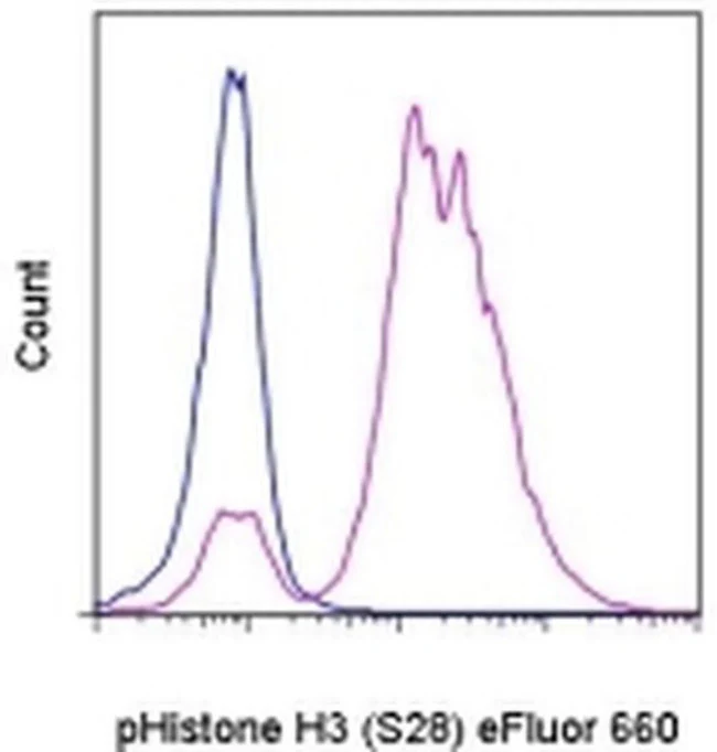
Applications Tested: This HTA28 antibody has been pre-titrated and tested by intracellular staining followed by flow cytometric analysis of nocodazole-treated HeLa cells. This can be used at 5 µL (0.25 µg) per test. A test is defined as the amount (µg) of antibody that will stain a cell sample in a final volume of 100 µL. Cell number should be determined empirically but can range from 10^5 to 10^8 cells/test.
Protocols: We recommend using Protocol A: Two-step protocol: intracellular (cytoplasmic) proteins or Protocol C: Two-step protocol: Fixation/Methanol. Protocol B: One-step protocol: intracellular (nuclear) proteins cannot be used. All Protocols can be found in the "Staining intracellular Antigens for Flow Cytometry Protocol" located in the BestProtocols® Section under the Resources tab online.
eFluor® 660 is a replacement for Alexa Fluor® 647. eFluor® 660 emits at 659 nm and is excited with the red laser (633 nm). Please make sure that your instrument is capable of detecting this fluorochrome.
Excitation: 633-647 nm; Emission: 668 nm; Laser: Red Laser.
Filtration: 0.2 µm post-manufacturing filtered.
For Research Use Only. Not for use in diagnostic procedures. Not for resale without express authorization.
Phospho-Histone H3 (Ser28) Monoclonal Antibody (HTA28), eFluor 660, eBioscience
PRODUCT DETAILS
Host: Rat
Isotype: IgG2a, kappa
Clonality: Monoclonal
Clone: HTA28
Format: eFluor 660
Reactivity: Hu, Ms
Application: Flow Cytometry
Tested Dilution: 5 µL (0.25 µg)/test
Concentration: 5 μL/Test
Storage: 4°C, store in dark, DO NOT FREEZE!
Formulation: PBS with BSA and 0.09% sodium azide; pH 7.2
Purification: Affinity chromatography
Data Sheet: TDS
Specific Information
Description: The HTA28 monoclonal antibody recognizes phosphorylated serine 28 of human, mouse, rat, bovine, and hamster histone H3. This 15 kDa protein is a component of eukaryotic chromatin that is involved in forming the nucleosome structure. Histone H3 can be phosphorylated at serine 10 and serine 28. Studies have demonstrated that phosphorylation at serine 28 is mediated by MSK1 following activation by the MAP kinase signaling pathway in response to tumor promoters (e.g., UV and EGF) and oncoproteins (e.g., c-Myc, c-Jun, and c-Fos). Histone H3 serine 28 phosphorylation has been linked to chromosome condensation during mitosis, cell transformation, and regulation of RNA polymerase III transcription machinery.
Applications Reported:This HTA28 antibody has been reported for use in intracellular staining followed by flow cytometric analysis.
Applications Tested: This HTA28 antibody has been pre-titrated and tested by intracellular staining followed by flow cytometric analysis of nocodazole-treated HeLa cells. This can be used at 5 µL (0.25 µg) per test. A test is defined as the amount (µg) of antibody that will stain a cell sample in a final volume of 100 µL. Cell number should be determined empirically but can range from 10^5 to 10^8 cells/test.
Protocols: We recommend using Protocol A: Two-step protocol: intracellular (cytoplasmic) proteins or Protocol C: Two-step protocol: Fixation/Methanol. Protocol B: One-step protocol: intracellular (nuclear) proteins cannot be used. All Protocols can be found in the "Staining intracellular Antigens for Flow Cytometry Protocol" located in the BestProtocols® Section under the Resources tab online.
eFluor® 660 is a replacement for Alexa Fluor® 647. eFluor® 660 emits at 659 nm and is excited with the red laser (633 nm). Please make sure that your instrument is capable of detecting this fluorochrome.
Excitation: 633-647 nm; Emission: 668 nm; Laser: Red Laser.
Filtration: 0.2 µm post-manufacturing filtered.
For Research Use Only. Not for use in diagnostic procedures. Not for resale without express authorization.
Product Information
Product Information
Shipping & Returns
Shipping & Returns
Description
PRODUCT DETAILS
Host: Rat
Isotype: IgG2a, kappa
Clonality: Monoclonal
Clone: HTA28
Format: eFluor 660
Reactivity: Hu, Ms
Application: Flow Cytometry
Tested Dilution: 5 µL (0.25 µg)/test
Concentration: 5 μL/Test
Storage: 4°C, store in dark, DO NOT FREEZE!
Formulation: PBS with BSA and 0.09% sodium azide; pH 7.2
Purification: Affinity chromatography
Data Sheet: TDS
Specific Information
Description: The HTA28 monoclonal antibody recognizes phosphorylated serine 28 of human, mouse, rat, bovine, and hamster histone H3. This 15 kDa protein is a component of eukaryotic chromatin that is involved in forming the nucleosome structure. Histone H3 can be phosphorylated at serine 10 and serine 28. Studies have demonstrated that phosphorylation at serine 28 is mediated by MSK1 following activation by the MAP kinase signaling pathway in response to tumor promoters (e.g., UV and EGF) and oncoproteins (e.g., c-Myc, c-Jun, and c-Fos). Histone H3 serine 28 phosphorylation has been linked to chromosome condensation during mitosis, cell transformation, and regulation of RNA polymerase III transcription machinery.
Applications Reported:This HTA28 antibody has been reported for use in intracellular staining followed by flow cytometric analysis.
Applications Tested: This HTA28 antibody has been pre-titrated and tested by intracellular staining followed by flow cytometric analysis of nocodazole-treated HeLa cells. This can be used at 5 µL (0.25 µg) per test. A test is defined as the amount (µg) of antibody that will stain a cell sample in a final volume of 100 µL. Cell number should be determined empirically but can range from 10^5 to 10^8 cells/test.
Protocols: We recommend using Protocol A: Two-step protocol: intracellular (cytoplasmic) proteins or Protocol C: Two-step protocol: Fixation/Methanol. Protocol B: One-step protocol: intracellular (nuclear) proteins cannot be used. All Protocols can be found in the "Staining intracellular Antigens for Flow Cytometry Protocol" located in the BestProtocols® Section under the Resources tab online.
eFluor® 660 is a replacement for Alexa Fluor® 647. eFluor® 660 emits at 659 nm and is excited with the red laser (633 nm). Please make sure that your instrument is capable of detecting this fluorochrome.
Excitation: 633-647 nm; Emission: 668 nm; Laser: Red Laser.
Filtration: 0.2 µm post-manufacturing filtered.
For Research Use Only. Not for use in diagnostic procedures. Not for resale without express authorization.


