Phospho-ERK1/2 (Thr202, Tyr204) Monoclonal Antibody (MILAN8R), APC, eBioscience
PRODUCT DETAILS
Host: Mouse
Isotype: IgG1, kappa
Clonality: Monoclonal
Clone: MILAN8R
Format: APC
Reactivity: Hu, Ms
Application: Flow Cytometry
Tested Dilution: 5 µL (0.03 µg)/test
Concentration: 5 μL/Test
Storage: 4°C, store in dark, DO NOT FREEZE!
Formulation: PBS with BSA and 0.09% sodium azide; pH 7.2
Purification: Affinity chromatography
Data Sheet: TDS
Specific Information
Description: This MILAN8R monoclonal antibody recognizes human and mouse extracellular signal-regulated kinases 1 and 2 (also known as ERK1/2, p44/p42, or MAPK3/1) when phosphorylated on T202/Y204. ERK1/2 belong to a family of conserved serine/threonine protein kinases known as mitogen-activated protein kinases (MAPKs) that are involved in many cellular programs such as proliferation, differentiation, motility, and survival. ERK1/2 signaling is activated in response to numerous extracellular stimuli including mitogens, growth factors, and cytokines. The primary activators of ERK1/2 are MEK1 and MEK2 which act by phosphorylating the activation loop residues T202/Y204 and T185/Y187 in ERK1 and ERK2, respectively. Several downstream targets of ERK1/2 have been identified, including p90RSK and the transcription factor Elk-1. ERK1/2 are negatively regulated by MAPK phosphatases, known as DUSPs or MKPs, as well as by chemical inhibitors of MEK including U0126 and PD98059. Disruption of the ERK pathway is common in many types of cancer.
Specificity of this MILAN8R clone was determined by ELISA, flow cytometry, and western blotting.
Applications Reported:This MILAN8R antibody has been reported for use in intracellular staining followed by flow cytometric analysis.
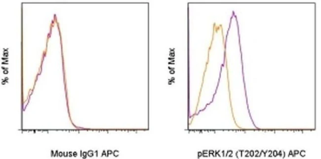
Applications Tested: This MILAN8R antibody has been pre-titrated and tested by intracellular staining and flow cytometric analysis of stimulated normal human peripheral blood cells. This can be used at 5 µL (0.03 µg) per test. A test is defined as the amount (µg) of antibody that will stain a cell sample in a final volume of 100 µL. Cell number should be determined empirically but can range from 10^5 to 10^8 cells/test.
Staining Protocol: We recommend using Protocol C: Two-step protocol: Fixation/Methanol. Protocol A: Two-step protocol: intracellular (cytoplasmic) proteins and Protocol B: One-step protocol: intracellular (nuclear) proteins cannot be used. All Protocols can be found in the Flow Cytometry Protocols: "Staining Intracellular Antigens for Flow Cytometry Protocol" located in the BestProtocols® Section under the Resources tab online.
Excitation: 633-647 nm; Emission: 660 nm; Laser: Red Laser.
Filtration: 0.2 µm post-manufacturing filtered.
For Research Use Only. Not for use in diagnostic procedures. Not for resale without express authorization.
Original: $439.00
-70%$439.00
$131.70Phospho-ERK1/2 (Thr202, Tyr204) Monoclonal Antibody (MILAN8R), APC, eBioscience
PRODUCT DETAILS
Host: Mouse
Isotype: IgG1, kappa
Clonality: Monoclonal
Clone: MILAN8R
Format: APC
Reactivity: Hu, Ms
Application: Flow Cytometry
Tested Dilution: 5 µL (0.03 µg)/test
Concentration: 5 μL/Test
Storage: 4°C, store in dark, DO NOT FREEZE!
Formulation: PBS with BSA and 0.09% sodium azide; pH 7.2
Purification: Affinity chromatography
Data Sheet: TDS
Specific Information
Description: This MILAN8R monoclonal antibody recognizes human and mouse extracellular signal-regulated kinases 1 and 2 (also known as ERK1/2, p44/p42, or MAPK3/1) when phosphorylated on T202/Y204. ERK1/2 belong to a family of conserved serine/threonine protein kinases known as mitogen-activated protein kinases (MAPKs) that are involved in many cellular programs such as proliferation, differentiation, motility, and survival. ERK1/2 signaling is activated in response to numerous extracellular stimuli including mitogens, growth factors, and cytokines. The primary activators of ERK1/2 are MEK1 and MEK2 which act by phosphorylating the activation loop residues T202/Y204 and T185/Y187 in ERK1 and ERK2, respectively. Several downstream targets of ERK1/2 have been identified, including p90RSK and the transcription factor Elk-1. ERK1/2 are negatively regulated by MAPK phosphatases, known as DUSPs or MKPs, as well as by chemical inhibitors of MEK including U0126 and PD98059. Disruption of the ERK pathway is common in many types of cancer.
Specificity of this MILAN8R clone was determined by ELISA, flow cytometry, and western blotting.
Applications Reported:This MILAN8R antibody has been reported for use in intracellular staining followed by flow cytometric analysis.
Applications Tested: This MILAN8R antibody has been pre-titrated and tested by intracellular staining and flow cytometric analysis of stimulated normal human peripheral blood cells. This can be used at 5 µL (0.03 µg) per test. A test is defined as the amount (µg) of antibody that will stain a cell sample in a final volume of 100 µL. Cell number should be determined empirically but can range from 10^5 to 10^8 cells/test.
Staining Protocol: We recommend using Protocol C: Two-step protocol: Fixation/Methanol. Protocol A: Two-step protocol: intracellular (cytoplasmic) proteins and Protocol B: One-step protocol: intracellular (nuclear) proteins cannot be used. All Protocols can be found in the Flow Cytometry Protocols: "Staining Intracellular Antigens for Flow Cytometry Protocol" located in the BestProtocols® Section under the Resources tab online.
Excitation: 633-647 nm; Emission: 660 nm; Laser: Red Laser.
Filtration: 0.2 µm post-manufacturing filtered.
For Research Use Only. Not for use in diagnostic procedures. Not for resale without express authorization.
Product Information
Product Information
Shipping & Returns
Shipping & Returns
Description
PRODUCT DETAILS
Host: Mouse
Isotype: IgG1, kappa
Clonality: Monoclonal
Clone: MILAN8R
Format: APC
Reactivity: Hu, Ms
Application: Flow Cytometry
Tested Dilution: 5 µL (0.03 µg)/test
Concentration: 5 μL/Test
Storage: 4°C, store in dark, DO NOT FREEZE!
Formulation: PBS with BSA and 0.09% sodium azide; pH 7.2
Purification: Affinity chromatography
Data Sheet: TDS
Specific Information
Description: This MILAN8R monoclonal antibody recognizes human and mouse extracellular signal-regulated kinases 1 and 2 (also known as ERK1/2, p44/p42, or MAPK3/1) when phosphorylated on T202/Y204. ERK1/2 belong to a family of conserved serine/threonine protein kinases known as mitogen-activated protein kinases (MAPKs) that are involved in many cellular programs such as proliferation, differentiation, motility, and survival. ERK1/2 signaling is activated in response to numerous extracellular stimuli including mitogens, growth factors, and cytokines. The primary activators of ERK1/2 are MEK1 and MEK2 which act by phosphorylating the activation loop residues T202/Y204 and T185/Y187 in ERK1 and ERK2, respectively. Several downstream targets of ERK1/2 have been identified, including p90RSK and the transcription factor Elk-1. ERK1/2 are negatively regulated by MAPK phosphatases, known as DUSPs or MKPs, as well as by chemical inhibitors of MEK including U0126 and PD98059. Disruption of the ERK pathway is common in many types of cancer.
Specificity of this MILAN8R clone was determined by ELISA, flow cytometry, and western blotting.
Applications Reported:This MILAN8R antibody has been reported for use in intracellular staining followed by flow cytometric analysis.
Applications Tested: This MILAN8R antibody has been pre-titrated and tested by intracellular staining and flow cytometric analysis of stimulated normal human peripheral blood cells. This can be used at 5 µL (0.03 µg) per test. A test is defined as the amount (µg) of antibody that will stain a cell sample in a final volume of 100 µL. Cell number should be determined empirically but can range from 10^5 to 10^8 cells/test.
Staining Protocol: We recommend using Protocol C: Two-step protocol: Fixation/Methanol. Protocol A: Two-step protocol: intracellular (cytoplasmic) proteins and Protocol B: One-step protocol: intracellular (nuclear) proteins cannot be used. All Protocols can be found in the Flow Cytometry Protocols: "Staining Intracellular Antigens for Flow Cytometry Protocol" located in the BestProtocols® Section under the Resources tab online.
Excitation: 633-647 nm; Emission: 660 nm; Laser: Red Laser.
Filtration: 0.2 µm post-manufacturing filtered.
For Research Use Only. Not for use in diagnostic procedures. Not for resale without express authorization.


