Phospho-CHK2 (Thr68) Monoclonal Antibody (ebchk2), PE, eBioscience
PRODUCT DETAILS
Host: Mouse
Isotype: IgG2b, kappa
Clonality: Monoclonal
Clone: ebchk2
Format: PE
Reactivity: Hu, Ms
Application: Flow Cytometry
Tested Dilution: 5 µL (0.125 µg)/test
Concentration: 5 μL/Test
Storage: 4°C, store in dark, DO NOT FREEZE!
Formulation: PBS with BSA and 0.09% sodium azide; pH 7.2
Purification: Affinity chromatography
Data Sheet: TDS
Specific Information
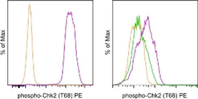
Description: This ebchk2 monoclonal antibody recognizes human and mouse Checkpoint Kinase 2 (Chk2) when phosphorylated on threonine 68 (T68). Chk2 is a cell cycle checkpoint regulator and tumor suppressor. Along with Chk1, it plays a critical role in the cellular response to DNA damage, and is particularly important during G2/M transition. Chk2 is activated via phosphorylation on T68 by the kinase ATM, which responds primarily to double-stranded DNA breaks. Upon activation, Chk2 functions include inhibition of Cdc25 family phosphatases and activation of p53, both of which prevent progression of the cell cycle. Abnormal regulation of the ATM-Chk2 pathway is often observed in cancer.
Applications Reported:This ebchk2 antibody has been reported for use in intracellular staining followed by flow cytometric analysis.
Applications Tested: This ebchk2 antibody has been pre-titrated and tested by intracellular staining followed by flow cytometric analysis of normal human peripheral blood cells. This can be used at 5 µL (0.125 µg) per test. A test is defined as the amount (µg) of antibody that will stain a cell sample in a final volume of 100 µL. Cell number should be determined empirically but can range from 10^5 to 10^8 cells/test.
Staining Protocol: All protocols work well for this monoclonal antibody. Use of Protocol A: Two-step protocol: intracellular (cytoplasmic) proteins allows for the greatest flexibility for detection of surface and intracellular (cytoplasmic) proteins. Use of Protocol B: One-step protocol: intracellular (nuclear) proteins is recommended for staining of transcription factors in conjunction with surface and phosphorylated intracellular (cytoplasmic) proteins. Protocol C: Two-step protocol: Fixation/Methanol allows for the greatest discrimination of phospho-specific signaling between unstimulated and stimulated samples, but with limitations on the ability to stain specific surface proteins (refer to "Clone Performance Following Fixation/Permeabilization" located in the BestProtocols Section under the Resources tab online). All Protocols can be found in the Flow Cytometry Protocols: "Staining Intracellular Antigens for Flow Cytometry Protocol" located in the BestProtocols® Section under the Resources tab online.
Excitation: 488-561 nm; Emission: 578 nm; Laser: Blue Laser, Green Laser, Yellow-Green Laser.
Filtration: 0.2 µm post-manufacturing filtered.
For Research Use Only. Not for use in diagnostic procedures. Not for resale without express authorization.
Phospho-CHK2 (Thr68) Monoclonal Antibody (ebchk2), PE, eBioscience
PRODUCT DETAILS
Host: Mouse
Isotype: IgG2b, kappa
Clonality: Monoclonal
Clone: ebchk2
Format: PE
Reactivity: Hu, Ms
Application: Flow Cytometry
Tested Dilution: 5 µL (0.125 µg)/test
Concentration: 5 μL/Test
Storage: 4°C, store in dark, DO NOT FREEZE!
Formulation: PBS with BSA and 0.09% sodium azide; pH 7.2
Purification: Affinity chromatography
Data Sheet: TDS
Specific Information
Description: This ebchk2 monoclonal antibody recognizes human and mouse Checkpoint Kinase 2 (Chk2) when phosphorylated on threonine 68 (T68). Chk2 is a cell cycle checkpoint regulator and tumor suppressor. Along with Chk1, it plays a critical role in the cellular response to DNA damage, and is particularly important during G2/M transition. Chk2 is activated via phosphorylation on T68 by the kinase ATM, which responds primarily to double-stranded DNA breaks. Upon activation, Chk2 functions include inhibition of Cdc25 family phosphatases and activation of p53, both of which prevent progression of the cell cycle. Abnormal regulation of the ATM-Chk2 pathway is often observed in cancer.
Applications Reported:This ebchk2 antibody has been reported for use in intracellular staining followed by flow cytometric analysis.
Applications Tested: This ebchk2 antibody has been pre-titrated and tested by intracellular staining followed by flow cytometric analysis of normal human peripheral blood cells. This can be used at 5 µL (0.125 µg) per test. A test is defined as the amount (µg) of antibody that will stain a cell sample in a final volume of 100 µL. Cell number should be determined empirically but can range from 10^5 to 10^8 cells/test.
Staining Protocol: All protocols work well for this monoclonal antibody. Use of Protocol A: Two-step protocol: intracellular (cytoplasmic) proteins allows for the greatest flexibility for detection of surface and intracellular (cytoplasmic) proteins. Use of Protocol B: One-step protocol: intracellular (nuclear) proteins is recommended for staining of transcription factors in conjunction with surface and phosphorylated intracellular (cytoplasmic) proteins. Protocol C: Two-step protocol: Fixation/Methanol allows for the greatest discrimination of phospho-specific signaling between unstimulated and stimulated samples, but with limitations on the ability to stain specific surface proteins (refer to "Clone Performance Following Fixation/Permeabilization" located in the BestProtocols Section under the Resources tab online). All Protocols can be found in the Flow Cytometry Protocols: "Staining Intracellular Antigens for Flow Cytometry Protocol" located in the BestProtocols® Section under the Resources tab online.
Excitation: 488-561 nm; Emission: 578 nm; Laser: Blue Laser, Green Laser, Yellow-Green Laser.
Filtration: 0.2 µm post-manufacturing filtered.
For Research Use Only. Not for use in diagnostic procedures. Not for resale without express authorization.
Product Information
Product Information
Shipping & Returns
Shipping & Returns
Description
PRODUCT DETAILS
Host: Mouse
Isotype: IgG2b, kappa
Clonality: Monoclonal
Clone: ebchk2
Format: PE
Reactivity: Hu, Ms
Application: Flow Cytometry
Tested Dilution: 5 µL (0.125 µg)/test
Concentration: 5 μL/Test
Storage: 4°C, store in dark, DO NOT FREEZE!
Formulation: PBS with BSA and 0.09% sodium azide; pH 7.2
Purification: Affinity chromatography
Data Sheet: TDS
Specific Information
Description: This ebchk2 monoclonal antibody recognizes human and mouse Checkpoint Kinase 2 (Chk2) when phosphorylated on threonine 68 (T68). Chk2 is a cell cycle checkpoint regulator and tumor suppressor. Along with Chk1, it plays a critical role in the cellular response to DNA damage, and is particularly important during G2/M transition. Chk2 is activated via phosphorylation on T68 by the kinase ATM, which responds primarily to double-stranded DNA breaks. Upon activation, Chk2 functions include inhibition of Cdc25 family phosphatases and activation of p53, both of which prevent progression of the cell cycle. Abnormal regulation of the ATM-Chk2 pathway is often observed in cancer.
Applications Reported:This ebchk2 antibody has been reported for use in intracellular staining followed by flow cytometric analysis.
Applications Tested: This ebchk2 antibody has been pre-titrated and tested by intracellular staining followed by flow cytometric analysis of normal human peripheral blood cells. This can be used at 5 µL (0.125 µg) per test. A test is defined as the amount (µg) of antibody that will stain a cell sample in a final volume of 100 µL. Cell number should be determined empirically but can range from 10^5 to 10^8 cells/test.
Staining Protocol: All protocols work well for this monoclonal antibody. Use of Protocol A: Two-step protocol: intracellular (cytoplasmic) proteins allows for the greatest flexibility for detection of surface and intracellular (cytoplasmic) proteins. Use of Protocol B: One-step protocol: intracellular (nuclear) proteins is recommended for staining of transcription factors in conjunction with surface and phosphorylated intracellular (cytoplasmic) proteins. Protocol C: Two-step protocol: Fixation/Methanol allows for the greatest discrimination of phospho-specific signaling between unstimulated and stimulated samples, but with limitations on the ability to stain specific surface proteins (refer to "Clone Performance Following Fixation/Permeabilization" located in the BestProtocols Section under the Resources tab online). All Protocols can be found in the Flow Cytometry Protocols: "Staining Intracellular Antigens for Flow Cytometry Protocol" located in the BestProtocols® Section under the Resources tab online.
Excitation: 488-561 nm; Emission: 578 nm; Laser: Blue Laser, Green Laser, Yellow-Green Laser.
Filtration: 0.2 µm post-manufacturing filtered.
For Research Use Only. Not for use in diagnostic procedures. Not for resale without express authorization.


