Nur77 Monoclonal Antibody (12.14), PE-Cyanine7, eBioscience
PRODUCT DETAILS
Host: Mouse
Isotype: IgG1, kappa
Clonality: Monoclonal
Clone: 12.14
Format: PE-Cyanine7
Reactivity: Ms
Application: Flow Cytometry
Tested Dilution: 0.25 µg/test
Concentration: 0.2 mg/mL
Storage: 4°C, store in dark, DO NOT FREEZE!
Formulation: PBS with 0.09% sodium azide; pH 7.2
Purification: Affinity chromatography
Data Sheet: TDS
Specific Information
Description: This 12.14 monoclonal antibody reacts with mouse Nur77 (also known as NR4A1, TR3, NGFI-B, or NAK1), an inducible orphan nuclear receptor. Expressed in thymocytes and T cell lines, Nur77 promotes apoptosis and plays a role in thymocyte negative selection. Additionally, Nur77 has been shown to be critical for steroid biosynthesis in Leydig cells as well as for the effects of dopamine. In addition, Nur77 has been shown to interact with FoxP3 in regulatory T cells. However, our results with this antibody do not correlate with this observation.
Applications Reported: This 12.14 antibody has been reported for use in intracellular staining followed by flow cytometric analysis.
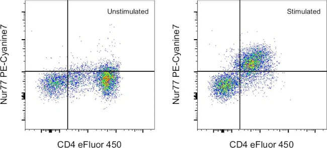
Applications Tested: This 12.14 antibody has been tested by intracellular staining followed by flow cytometric analysis of stimulated mouse thymocytes using the Foxp3/Transcription Factor Staining Buffer Set (Product # 00-5523-00) and protocol. Please refer to "Staining Intracellular Antigens for Flow Cytometry, Protocol B: One step protocol for intracellular (nuclear) proteins" located at www.thermofisher.com/flowprotocols . This may be used at less than or equal to 0.25 µg per test. A test is defined as the amount (µg) of antibody that will stain a cell sample in a final volume of 100 µL. Cell number should be determined empirically but can range from 10^5 to 10^8 cells/test. It is recommended that the antibody be carefully titrated for optimal performance in the assay of interest.
Light sensitivity: This tandem dye is sensitive to photo-induced oxidation. Please protect this vial and stained samples from light.
Fixation: Samples can be stored in IC Fixation Buffer (Product # 00-8222-49) (100 µL of cell sample + 100 µL of IC Fixation Buffer) or 1-step Fix/Lyse Solution (Product # 00-5333-57) for up to 3 days in the dark at 4°C with minimal impact on brightness and FRET efficiency/compensation. Some generalizations regarding fluorophore performance after fixation can be made, but clone specific performance should be determined empirically.
Excitation: 488-561 nm; Emission: 775 nm; Laser: Blue Laser, Green Laser, Yellow-Green Laser.
For Research Use Only. Not for use in diagnostic procedures. Not for resale without express authorization.
Original: $454.00
-70%$454.00
$136.20Nur77 Monoclonal Antibody (12.14), PE-Cyanine7, eBioscience
PRODUCT DETAILS
Host: Mouse
Isotype: IgG1, kappa
Clonality: Monoclonal
Clone: 12.14
Format: PE-Cyanine7
Reactivity: Ms
Application: Flow Cytometry
Tested Dilution: 0.25 µg/test
Concentration: 0.2 mg/mL
Storage: 4°C, store in dark, DO NOT FREEZE!
Formulation: PBS with 0.09% sodium azide; pH 7.2
Purification: Affinity chromatography
Data Sheet: TDS
Specific Information
Description: This 12.14 monoclonal antibody reacts with mouse Nur77 (also known as NR4A1, TR3, NGFI-B, or NAK1), an inducible orphan nuclear receptor. Expressed in thymocytes and T cell lines, Nur77 promotes apoptosis and plays a role in thymocyte negative selection. Additionally, Nur77 has been shown to be critical for steroid biosynthesis in Leydig cells as well as for the effects of dopamine. In addition, Nur77 has been shown to interact with FoxP3 in regulatory T cells. However, our results with this antibody do not correlate with this observation.
Applications Reported: This 12.14 antibody has been reported for use in intracellular staining followed by flow cytometric analysis.
Applications Tested: This 12.14 antibody has been tested by intracellular staining followed by flow cytometric analysis of stimulated mouse thymocytes using the Foxp3/Transcription Factor Staining Buffer Set (Product # 00-5523-00) and protocol. Please refer to "Staining Intracellular Antigens for Flow Cytometry, Protocol B: One step protocol for intracellular (nuclear) proteins" located at www.thermofisher.com/flowprotocols . This may be used at less than or equal to 0.25 µg per test. A test is defined as the amount (µg) of antibody that will stain a cell sample in a final volume of 100 µL. Cell number should be determined empirically but can range from 10^5 to 10^8 cells/test. It is recommended that the antibody be carefully titrated for optimal performance in the assay of interest.
Light sensitivity: This tandem dye is sensitive to photo-induced oxidation. Please protect this vial and stained samples from light.
Fixation: Samples can be stored in IC Fixation Buffer (Product # 00-8222-49) (100 µL of cell sample + 100 µL of IC Fixation Buffer) or 1-step Fix/Lyse Solution (Product # 00-5333-57) for up to 3 days in the dark at 4°C with minimal impact on brightness and FRET efficiency/compensation. Some generalizations regarding fluorophore performance after fixation can be made, but clone specific performance should be determined empirically.
Excitation: 488-561 nm; Emission: 775 nm; Laser: Blue Laser, Green Laser, Yellow-Green Laser.
For Research Use Only. Not for use in diagnostic procedures. Not for resale without express authorization.
Product Information
Product Information
Shipping & Returns
Shipping & Returns
Description
PRODUCT DETAILS
Host: Mouse
Isotype: IgG1, kappa
Clonality: Monoclonal
Clone: 12.14
Format: PE-Cyanine7
Reactivity: Ms
Application: Flow Cytometry
Tested Dilution: 0.25 µg/test
Concentration: 0.2 mg/mL
Storage: 4°C, store in dark, DO NOT FREEZE!
Formulation: PBS with 0.09% sodium azide; pH 7.2
Purification: Affinity chromatography
Data Sheet: TDS
Specific Information
Description: This 12.14 monoclonal antibody reacts with mouse Nur77 (also known as NR4A1, TR3, NGFI-B, or NAK1), an inducible orphan nuclear receptor. Expressed in thymocytes and T cell lines, Nur77 promotes apoptosis and plays a role in thymocyte negative selection. Additionally, Nur77 has been shown to be critical for steroid biosynthesis in Leydig cells as well as for the effects of dopamine. In addition, Nur77 has been shown to interact with FoxP3 in regulatory T cells. However, our results with this antibody do not correlate with this observation.
Applications Reported: This 12.14 antibody has been reported for use in intracellular staining followed by flow cytometric analysis.
Applications Tested: This 12.14 antibody has been tested by intracellular staining followed by flow cytometric analysis of stimulated mouse thymocytes using the Foxp3/Transcription Factor Staining Buffer Set (Product # 00-5523-00) and protocol. Please refer to "Staining Intracellular Antigens for Flow Cytometry, Protocol B: One step protocol for intracellular (nuclear) proteins" located at www.thermofisher.com/flowprotocols . This may be used at less than or equal to 0.25 µg per test. A test is defined as the amount (µg) of antibody that will stain a cell sample in a final volume of 100 µL. Cell number should be determined empirically but can range from 10^5 to 10^8 cells/test. It is recommended that the antibody be carefully titrated for optimal performance in the assay of interest.
Light sensitivity: This tandem dye is sensitive to photo-induced oxidation. Please protect this vial and stained samples from light.
Fixation: Samples can be stored in IC Fixation Buffer (Product # 00-8222-49) (100 µL of cell sample + 100 µL of IC Fixation Buffer) or 1-step Fix/Lyse Solution (Product # 00-5333-57) for up to 3 days in the dark at 4°C with minimal impact on brightness and FRET efficiency/compensation. Some generalizations regarding fluorophore performance after fixation can be made, but clone specific performance should be determined empirically.
Excitation: 488-561 nm; Emission: 775 nm; Laser: Blue Laser, Green Laser, Yellow-Green Laser.
For Research Use Only. Not for use in diagnostic procedures. Not for resale without express authorization.


