NOTCH2 Monoclonal Antibody (16F11), PE, eBioscience
PRODUCT DETAILS
Host: Rat
Isotype: IgG1, kappa
Clonality: Monoclonal
Clone: 16F11
Format: PE
Reactivity: Hu, Ms
Application: Flow Cytometry
Tested Dilution: 1 µg/test
Concentration: 0.2 mg/mL
Storage: 4°C, store in dark, DO NOT FREEZE!
Formulation: PBS with 0.09% sodium azide; pH 7.2
Purification: Affinity chromatography
Data Sheet: TDS
Specific Information
Description: This 16F11 monoclonal antibody reacts with the extracellular domain of human and mouse Notch2, one of four members of the Notch family of receptors. Notch receptors are 300-kDa single-pass transmembrane proteins. While the extracellular domain contains numerous epidermal growth factor-like repeats for ligand binding, the intracellular domain is involved in cell signaling. Upon binding its membrane-bound ligand (either Delta or Jagged), the Notch receptor undergoes proteolytic cleavage, first by ADAM-family metalloproteases and then by gamma-secretase. The second cleavage event releases the Notch intracellular domain (NICD), which subsequently translocates into the nucleus, heterodimerizes with the DNA-binding protein RBP-J, recruits co-activator molecules, and ultimately activates transcription.
Notch2 is expressed on thymocytes (i.e., DN1 and DN2 subsets), activated peripheral T cells, and bone marrow. Moreover, expression of Notch2 has been reported to be altered in various cancers and lymphomas. In addition to its role in stem cell hematopoiesis, Notch2 plays a pivotal role in marginal zone B cell development. Notch2 also interacts with phosphorylated CREB1 to induce granzyme B transcription in cytotoxic T cells.
Applications Reported: This 16F11 antibody has been reported for use in flow cytometric analysis.
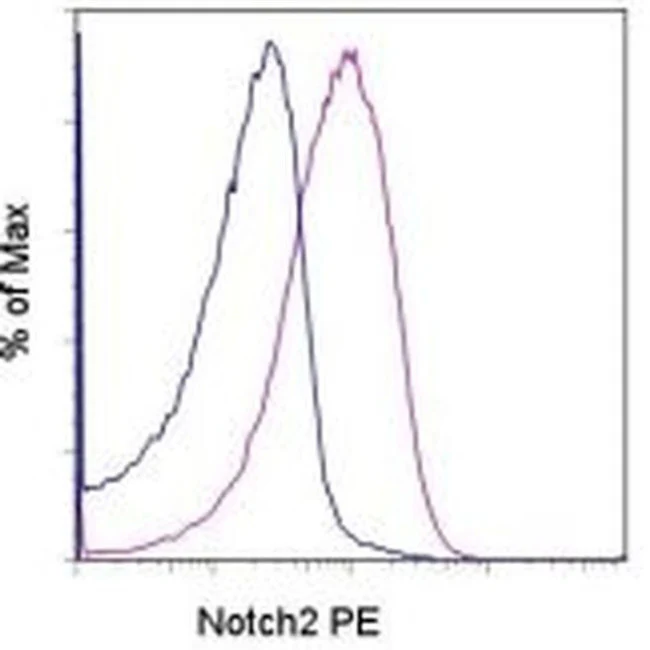
Applications Tested: This 16F11 antibody has been tested by flow cytometric analysis of 24-hour anti-CD3-stimulated mouse splenocytes. This can be used at less than or equal to 1 µg per test. A test is defined as the amount (µg) of antibody that will stain a cell sample in a final volume of 100 µL. Cell number should be determined empirically but can range from 10^5 to 10^8 cells/test. It is recommended that the antibody be carefully titrated for optimal performance in the assay of interest.
Excitation: 488-561 nm; Emission: 578 nm; Laser: Blue Laser, Green Laser, Yellow-Green Laser.
Filtration: 0.2 µm post-manufacturing filtered.
For Research Use Only. Not for use in diagnostic procedures. Not for resale without express authorization.
Original: $390.00
-70%$390.00
$117.00NOTCH2 Monoclonal Antibody (16F11), PE, eBioscience
PRODUCT DETAILS
Host: Rat
Isotype: IgG1, kappa
Clonality: Monoclonal
Clone: 16F11
Format: PE
Reactivity: Hu, Ms
Application: Flow Cytometry
Tested Dilution: 1 µg/test
Concentration: 0.2 mg/mL
Storage: 4°C, store in dark, DO NOT FREEZE!
Formulation: PBS with 0.09% sodium azide; pH 7.2
Purification: Affinity chromatography
Data Sheet: TDS
Specific Information
Description: This 16F11 monoclonal antibody reacts with the extracellular domain of human and mouse Notch2, one of four members of the Notch family of receptors. Notch receptors are 300-kDa single-pass transmembrane proteins. While the extracellular domain contains numerous epidermal growth factor-like repeats for ligand binding, the intracellular domain is involved in cell signaling. Upon binding its membrane-bound ligand (either Delta or Jagged), the Notch receptor undergoes proteolytic cleavage, first by ADAM-family metalloproteases and then by gamma-secretase. The second cleavage event releases the Notch intracellular domain (NICD), which subsequently translocates into the nucleus, heterodimerizes with the DNA-binding protein RBP-J, recruits co-activator molecules, and ultimately activates transcription.
Notch2 is expressed on thymocytes (i.e., DN1 and DN2 subsets), activated peripheral T cells, and bone marrow. Moreover, expression of Notch2 has been reported to be altered in various cancers and lymphomas. In addition to its role in stem cell hematopoiesis, Notch2 plays a pivotal role in marginal zone B cell development. Notch2 also interacts with phosphorylated CREB1 to induce granzyme B transcription in cytotoxic T cells.
Applications Reported: This 16F11 antibody has been reported for use in flow cytometric analysis.
Applications Tested: This 16F11 antibody has been tested by flow cytometric analysis of 24-hour anti-CD3-stimulated mouse splenocytes. This can be used at less than or equal to 1 µg per test. A test is defined as the amount (µg) of antibody that will stain a cell sample in a final volume of 100 µL. Cell number should be determined empirically but can range from 10^5 to 10^8 cells/test. It is recommended that the antibody be carefully titrated for optimal performance in the assay of interest.
Excitation: 488-561 nm; Emission: 578 nm; Laser: Blue Laser, Green Laser, Yellow-Green Laser.
Filtration: 0.2 µm post-manufacturing filtered.
For Research Use Only. Not for use in diagnostic procedures. Not for resale without express authorization.
Product Information
Product Information
Shipping & Returns
Shipping & Returns
Description
PRODUCT DETAILS
Host: Rat
Isotype: IgG1, kappa
Clonality: Monoclonal
Clone: 16F11
Format: PE
Reactivity: Hu, Ms
Application: Flow Cytometry
Tested Dilution: 1 µg/test
Concentration: 0.2 mg/mL
Storage: 4°C, store in dark, DO NOT FREEZE!
Formulation: PBS with 0.09% sodium azide; pH 7.2
Purification: Affinity chromatography
Data Sheet: TDS
Specific Information
Description: This 16F11 monoclonal antibody reacts with the extracellular domain of human and mouse Notch2, one of four members of the Notch family of receptors. Notch receptors are 300-kDa single-pass transmembrane proteins. While the extracellular domain contains numerous epidermal growth factor-like repeats for ligand binding, the intracellular domain is involved in cell signaling. Upon binding its membrane-bound ligand (either Delta or Jagged), the Notch receptor undergoes proteolytic cleavage, first by ADAM-family metalloproteases and then by gamma-secretase. The second cleavage event releases the Notch intracellular domain (NICD), which subsequently translocates into the nucleus, heterodimerizes with the DNA-binding protein RBP-J, recruits co-activator molecules, and ultimately activates transcription.
Notch2 is expressed on thymocytes (i.e., DN1 and DN2 subsets), activated peripheral T cells, and bone marrow. Moreover, expression of Notch2 has been reported to be altered in various cancers and lymphomas. In addition to its role in stem cell hematopoiesis, Notch2 plays a pivotal role in marginal zone B cell development. Notch2 also interacts with phosphorylated CREB1 to induce granzyme B transcription in cytotoxic T cells.
Applications Reported: This 16F11 antibody has been reported for use in flow cytometric analysis.
Applications Tested: This 16F11 antibody has been tested by flow cytometric analysis of 24-hour anti-CD3-stimulated mouse splenocytes. This can be used at less than or equal to 1 µg per test. A test is defined as the amount (µg) of antibody that will stain a cell sample in a final volume of 100 µL. Cell number should be determined empirically but can range from 10^5 to 10^8 cells/test. It is recommended that the antibody be carefully titrated for optimal performance in the assay of interest.
Excitation: 488-561 nm; Emission: 578 nm; Laser: Blue Laser, Green Laser, Yellow-Green Laser.
Filtration: 0.2 µm post-manufacturing filtered.
For Research Use Only. Not for use in diagnostic procedures. Not for resale without express authorization.


