NOTCH1 Monoclonal Antibody (mN1A), PE, eBioscience
PRODUCT DETAILS
Host: Mouse
Isotype: IgG1, kappa
Clonality: Monoclonal
Clone: mN1A
Format: PE
Reactivity: Hu, Ms
Application: Flow Cytometry
Tested Dilution: 0.25 µg/test
Concentration: 0.2 mg/mL
Storage: 4°C, store in dark, DO NOT FREEZE!
Formulation: PBS with 0.09% sodium azide; pH 7.2
Purification: Affinity chromatography
Data Sheet: TDS
Specific Information
Description: The Notch family of transmembrane receptors controls cell-fate decisions during the development of many organs in a wide variety of species. After binding its ligand, the Notch receptor is cleaved in its transmembrane domain, and the resulting intracellular domain dissociates from the membrane and translocates to the nucleus, where it is able to suppress the expression of lineage-specific genes by interacting with transcriptional repressors. The mN1A antibody reacts with the intracellular domain of mouse and human Notch1, but not with Notch2, 3, or 4. The mN1A antibody has a low affinity for the full-length (unprocessed or heterodimeric cell surface) forms of Notch1. In the mouse, Notch mRNA is expressed in mouse hematopoietic cells of the fetal liver and adult thymus and bone marrow. In the thymus, Notch1 protein is detected in CD4-CD8- (double-negative) and CD4-CD8+ (single-positive) thymocytes. Studies of Notch1-transgenic cells and Notch1-null mice indicate that the receptor is involved in the regulation of lymphopoiesis and myelopoiesis.
Applications Reported: This mN1A antibody has been reported for use in intracellular staining followed by flow cytometric analysis.
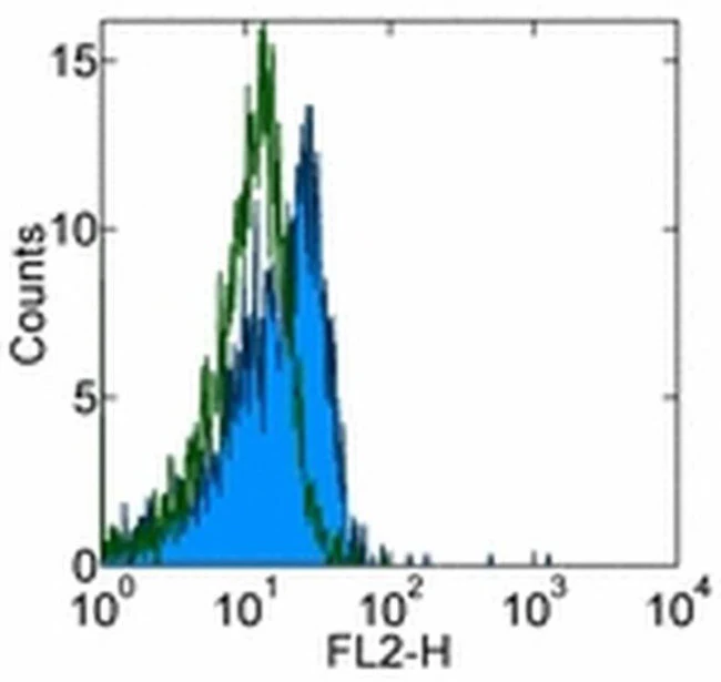
Applications Tested: This mN1A antibody has been tested by intracellular staining and flow cytometric analysis of mouse thymocytes using the Intracellular Fixation & Permeabilization Buffer Set (Product # 88-8824-00)and protocol. Please refer to BestProtocols®: Protocol A: Two step protocol for (cytoplasmic) intracellular proteins located under the Resources Tab online. This can be used at less than or equal to 0.25 µg per test. A test is defined as the amount (µg) of antibody that will stain a cell sample in a final volume of 100 µL. Cell number should be determined empirically but can range from 10^5 to 10^8 cells/test. It is recommended that the antibody be carefully titrated for optimal performance in the assay of interest.
Excitation: 488-561 nm; Emission: 578 nm; Laser: Blue Laser, Green Laser, Yellow-Green Laser.
Filtration: 0.2 µm post-manufacturing filtered.
For Research Use Only. Not for use in diagnostic procedures. Not for resale without express authorization.
Original: $266.00
-70%$266.00
$79.80NOTCH1 Monoclonal Antibody (mN1A), PE, eBioscience
PRODUCT DETAILS
Host: Mouse
Isotype: IgG1, kappa
Clonality: Monoclonal
Clone: mN1A
Format: PE
Reactivity: Hu, Ms
Application: Flow Cytometry
Tested Dilution: 0.25 µg/test
Concentration: 0.2 mg/mL
Storage: 4°C, store in dark, DO NOT FREEZE!
Formulation: PBS with 0.09% sodium azide; pH 7.2
Purification: Affinity chromatography
Data Sheet: TDS
Specific Information
Description: The Notch family of transmembrane receptors controls cell-fate decisions during the development of many organs in a wide variety of species. After binding its ligand, the Notch receptor is cleaved in its transmembrane domain, and the resulting intracellular domain dissociates from the membrane and translocates to the nucleus, where it is able to suppress the expression of lineage-specific genes by interacting with transcriptional repressors. The mN1A antibody reacts with the intracellular domain of mouse and human Notch1, but not with Notch2, 3, or 4. The mN1A antibody has a low affinity for the full-length (unprocessed or heterodimeric cell surface) forms of Notch1. In the mouse, Notch mRNA is expressed in mouse hematopoietic cells of the fetal liver and adult thymus and bone marrow. In the thymus, Notch1 protein is detected in CD4-CD8- (double-negative) and CD4-CD8+ (single-positive) thymocytes. Studies of Notch1-transgenic cells and Notch1-null mice indicate that the receptor is involved in the regulation of lymphopoiesis and myelopoiesis.
Applications Reported: This mN1A antibody has been reported for use in intracellular staining followed by flow cytometric analysis.
Applications Tested: This mN1A antibody has been tested by intracellular staining and flow cytometric analysis of mouse thymocytes using the Intracellular Fixation & Permeabilization Buffer Set (Product # 88-8824-00)and protocol. Please refer to BestProtocols®: Protocol A: Two step protocol for (cytoplasmic) intracellular proteins located under the Resources Tab online. This can be used at less than or equal to 0.25 µg per test. A test is defined as the amount (µg) of antibody that will stain a cell sample in a final volume of 100 µL. Cell number should be determined empirically but can range from 10^5 to 10^8 cells/test. It is recommended that the antibody be carefully titrated for optimal performance in the assay of interest.
Excitation: 488-561 nm; Emission: 578 nm; Laser: Blue Laser, Green Laser, Yellow-Green Laser.
Filtration: 0.2 µm post-manufacturing filtered.
For Research Use Only. Not for use in diagnostic procedures. Not for resale without express authorization.
Product Information
Product Information
Shipping & Returns
Shipping & Returns
Description
PRODUCT DETAILS
Host: Mouse
Isotype: IgG1, kappa
Clonality: Monoclonal
Clone: mN1A
Format: PE
Reactivity: Hu, Ms
Application: Flow Cytometry
Tested Dilution: 0.25 µg/test
Concentration: 0.2 mg/mL
Storage: 4°C, store in dark, DO NOT FREEZE!
Formulation: PBS with 0.09% sodium azide; pH 7.2
Purification: Affinity chromatography
Data Sheet: TDS
Specific Information
Description: The Notch family of transmembrane receptors controls cell-fate decisions during the development of many organs in a wide variety of species. After binding its ligand, the Notch receptor is cleaved in its transmembrane domain, and the resulting intracellular domain dissociates from the membrane and translocates to the nucleus, where it is able to suppress the expression of lineage-specific genes by interacting with transcriptional repressors. The mN1A antibody reacts with the intracellular domain of mouse and human Notch1, but not with Notch2, 3, or 4. The mN1A antibody has a low affinity for the full-length (unprocessed or heterodimeric cell surface) forms of Notch1. In the mouse, Notch mRNA is expressed in mouse hematopoietic cells of the fetal liver and adult thymus and bone marrow. In the thymus, Notch1 protein is detected in CD4-CD8- (double-negative) and CD4-CD8+ (single-positive) thymocytes. Studies of Notch1-transgenic cells and Notch1-null mice indicate that the receptor is involved in the regulation of lymphopoiesis and myelopoiesis.
Applications Reported: This mN1A antibody has been reported for use in intracellular staining followed by flow cytometric analysis.
Applications Tested: This mN1A antibody has been tested by intracellular staining and flow cytometric analysis of mouse thymocytes using the Intracellular Fixation & Permeabilization Buffer Set (Product # 88-8824-00)and protocol. Please refer to BestProtocols®: Protocol A: Two step protocol for (cytoplasmic) intracellular proteins located under the Resources Tab online. This can be used at less than or equal to 0.25 µg per test. A test is defined as the amount (µg) of antibody that will stain a cell sample in a final volume of 100 µL. Cell number should be determined empirically but can range from 10^5 to 10^8 cells/test. It is recommended that the antibody be carefully titrated for optimal performance in the assay of interest.
Excitation: 488-561 nm; Emission: 578 nm; Laser: Blue Laser, Green Laser, Yellow-Green Laser.
Filtration: 0.2 µm post-manufacturing filtered.
For Research Use Only. Not for use in diagnostic procedures. Not for resale without express authorization.


