MAVS Monoclonal Antibody (ABM28H9), APC, eBioscience
PRODUCT DETAILS
Host: Mouse
Isotype: IgG2b, kappa
Clonality: Monoclonal
Clone: ABM28H9
Format: APC
Reactivity: Hu
Application: Flow Cytometry
Tested Dilution: 5 µL (0.06 µg)/test
Concentration: 5 μL/Test
Storage: 4°C, store in dark, DO NOT FREEZE!
Formulation: PBS with BSA and 0.09% sodium azide; pH 7.2
Purification: Affinity chromatography
Data Sheet: TDS
Specific Information
Description: This ABM28H9 monoclonal antibody recognizes human MAVS, a mitochondrial membrane protein also known as VISA, CARDIF or IPS-1.
Applications Reported: This ABM18H9 antibody has been reported for use in intracellular staining followed by flow cytometric analysis.
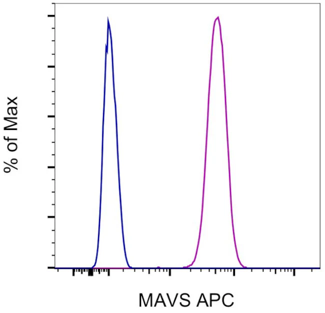
Applications Tested: This ABM18H9 antibody has been pre-diluted and tested by intracellular staining followed by flow cytometric analysis of HEL cells using the Intracellular Fixation & Permeabilization Buffer Set (Product # 88-8824-00) and protocol. Please refer to "Staining Intracellular Antigens for Flow Cytometry, Protocol A: Two step protocol for intracellular (cytoplasmic) proteins" located at www.thermofisher.com/flowprotocols . This may be used at 5 µL (0.06 µg) per test. A test is defined as the amount (µg) of antibody that will stain a cell sample in a final volume of 100 µL. Cell number should be determined empirically but can range from 10^5 to 10^8 cells/test.
Excitation: 633-647 nm; Emission: 660 nm; Laser: Red Laser.
For Research Use Only. Not for use in diagnostic procedures. Not for resale without express authorization.
MAVS Monoclonal Antibody (ABM28H9), APC, eBioscience
PRODUCT DETAILS
Host: Mouse
Isotype: IgG2b, kappa
Clonality: Monoclonal
Clone: ABM28H9
Format: APC
Reactivity: Hu
Application: Flow Cytometry
Tested Dilution: 5 µL (0.06 µg)/test
Concentration: 5 μL/Test
Storage: 4°C, store in dark, DO NOT FREEZE!
Formulation: PBS with BSA and 0.09% sodium azide; pH 7.2
Purification: Affinity chromatography
Data Sheet: TDS
Specific Information
Description: This ABM28H9 monoclonal antibody recognizes human MAVS, a mitochondrial membrane protein also known as VISA, CARDIF or IPS-1.
Applications Reported: This ABM18H9 antibody has been reported for use in intracellular staining followed by flow cytometric analysis.
Applications Tested: This ABM18H9 antibody has been pre-diluted and tested by intracellular staining followed by flow cytometric analysis of HEL cells using the Intracellular Fixation & Permeabilization Buffer Set (Product # 88-8824-00) and protocol. Please refer to "Staining Intracellular Antigens for Flow Cytometry, Protocol A: Two step protocol for intracellular (cytoplasmic) proteins" located at www.thermofisher.com/flowprotocols . This may be used at 5 µL (0.06 µg) per test. A test is defined as the amount (µg) of antibody that will stain a cell sample in a final volume of 100 µL. Cell number should be determined empirically but can range from 10^5 to 10^8 cells/test.
Excitation: 633-647 nm; Emission: 660 nm; Laser: Red Laser.
For Research Use Only. Not for use in diagnostic procedures. Not for resale without express authorization.
Product Information
Product Information
Shipping & Returns
Shipping & Returns
Description
PRODUCT DETAILS
Host: Mouse
Isotype: IgG2b, kappa
Clonality: Monoclonal
Clone: ABM28H9
Format: APC
Reactivity: Hu
Application: Flow Cytometry
Tested Dilution: 5 µL (0.06 µg)/test
Concentration: 5 μL/Test
Storage: 4°C, store in dark, DO NOT FREEZE!
Formulation: PBS with BSA and 0.09% sodium azide; pH 7.2
Purification: Affinity chromatography
Data Sheet: TDS
Specific Information
Description: This ABM28H9 monoclonal antibody recognizes human MAVS, a mitochondrial membrane protein also known as VISA, CARDIF or IPS-1.
Applications Reported: This ABM18H9 antibody has been reported for use in intracellular staining followed by flow cytometric analysis.
Applications Tested: This ABM18H9 antibody has been pre-diluted and tested by intracellular staining followed by flow cytometric analysis of HEL cells using the Intracellular Fixation & Permeabilization Buffer Set (Product # 88-8824-00) and protocol. Please refer to "Staining Intracellular Antigens for Flow Cytometry, Protocol A: Two step protocol for intracellular (cytoplasmic) proteins" located at www.thermofisher.com/flowprotocols . This may be used at 5 µL (0.06 µg) per test. A test is defined as the amount (µg) of antibody that will stain a cell sample in a final volume of 100 µL. Cell number should be determined empirically but can range from 10^5 to 10^8 cells/test.
Excitation: 633-647 nm; Emission: 660 nm; Laser: Red Laser.
For Research Use Only. Not for use in diagnostic procedures. Not for resale without express authorization.


