LYVE1 Monoclonal Antibody (ALY7), Biotin, eBioscience
PRODUCT DETAILS
Host: Rat
Isotype: IgG1, kappa
Clonality: Monoclonal
Clone: ALY7
Format: Biotin
Reactivity: Ms
Application: Flow Cytometry
Tested Dilution: 0.125 µg/test
Concentration: 0.5 mg/mL
Storage: 4°C, store in dark, DO NOT FREEZE!
Formulation: PBS with 0.09% sodium azide; pH 7.2
Purification: Affinity chromatography
Data Sheet: TDS
Specific Information
Description: The monoclonal antibody ALY7 recognizes mouse LYVE-1, a transmembrane glycoprotein with similarity to CD44. The extracellular domain contains a conserved hyaluronan binding domain also found in CD44. Expression is found on lymphatic and liver endothelial cells and some populations of macrophages. The lymphatic system is responsible for transporting proteins and cells (especially dendritic cells) to tissues throughout the body, thereby acting as immune surveyors. LYVE-1 is one characteristic protein, along with podoplanin, PROX-1, Tie-2 and VEGFR-3, that is expressed on lymphatic endothelial cells (LECS). The ligand for LYVE-1 is hyaluronan, a large mucopolysaccharide. Although LYVE-1 can bind hyaluronan in vitro, the site for ligand binding in vivo is masked by sialyated O-linked glycan chains. It is postulated that binding to ligand requires modification/unmasking to expose the binding site. The development and remodeling of the endothelium after injury is an area of extensive study. When transplanted, hematopoietic stem cells (HSCs) can give rise to LECs that integrate into the endothelium in normal and metastatic tissue.
Applications Reported: This ALY7 antibody has been reported for use in flow cytometric analysis, and immunohistology staining of frozen tissue sections.
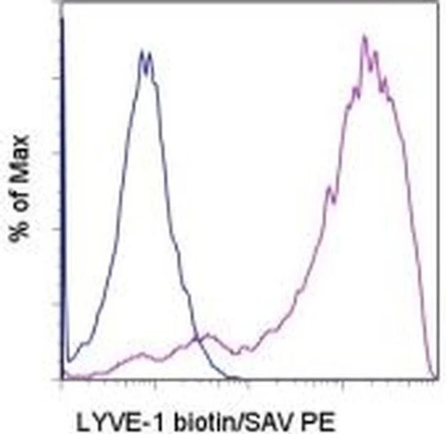
Applications Tested: This ALY7 antibody has been tested by flow cytometric analysis of LYVE-1/GFP co-transfected cells. This can be used at less than or equal to 0.125 µg per test. A test is defined as the amount (µg) of antibody that will stain a cell sample in a final volume of 100 µL. Cell number should be determined empirically but can range from 10^5 to 10^8 cells/test. It is recommended that the antibody be carefully titrated for optimal performance in the assay of interest.
Filtration: 0.2 µm post-manufacturing filtered.
For Research Use Only. Not for use in diagnostic procedures. Not for resale without express authorization.
LYVE1 Monoclonal Antibody (ALY7), Biotin, eBioscience
PRODUCT DETAILS
Host: Rat
Isotype: IgG1, kappa
Clonality: Monoclonal
Clone: ALY7
Format: Biotin
Reactivity: Ms
Application: Flow Cytometry
Tested Dilution: 0.125 µg/test
Concentration: 0.5 mg/mL
Storage: 4°C, store in dark, DO NOT FREEZE!
Formulation: PBS with 0.09% sodium azide; pH 7.2
Purification: Affinity chromatography
Data Sheet: TDS
Specific Information
Description: The monoclonal antibody ALY7 recognizes mouse LYVE-1, a transmembrane glycoprotein with similarity to CD44. The extracellular domain contains a conserved hyaluronan binding domain also found in CD44. Expression is found on lymphatic and liver endothelial cells and some populations of macrophages. The lymphatic system is responsible for transporting proteins and cells (especially dendritic cells) to tissues throughout the body, thereby acting as immune surveyors. LYVE-1 is one characteristic protein, along with podoplanin, PROX-1, Tie-2 and VEGFR-3, that is expressed on lymphatic endothelial cells (LECS). The ligand for LYVE-1 is hyaluronan, a large mucopolysaccharide. Although LYVE-1 can bind hyaluronan in vitro, the site for ligand binding in vivo is masked by sialyated O-linked glycan chains. It is postulated that binding to ligand requires modification/unmasking to expose the binding site. The development and remodeling of the endothelium after injury is an area of extensive study. When transplanted, hematopoietic stem cells (HSCs) can give rise to LECs that integrate into the endothelium in normal and metastatic tissue.
Applications Reported: This ALY7 antibody has been reported for use in flow cytometric analysis, and immunohistology staining of frozen tissue sections.
Applications Tested: This ALY7 antibody has been tested by flow cytometric analysis of LYVE-1/GFP co-transfected cells. This can be used at less than or equal to 0.125 µg per test. A test is defined as the amount (µg) of antibody that will stain a cell sample in a final volume of 100 µL. Cell number should be determined empirically but can range from 10^5 to 10^8 cells/test. It is recommended that the antibody be carefully titrated for optimal performance in the assay of interest.
Filtration: 0.2 µm post-manufacturing filtered.
For Research Use Only. Not for use in diagnostic procedures. Not for resale without express authorization.
Product Information
Product Information
Shipping & Returns
Shipping & Returns
Description
PRODUCT DETAILS
Host: Rat
Isotype: IgG1, kappa
Clonality: Monoclonal
Clone: ALY7
Format: Biotin
Reactivity: Ms
Application: Flow Cytometry
Tested Dilution: 0.125 µg/test
Concentration: 0.5 mg/mL
Storage: 4°C, store in dark, DO NOT FREEZE!
Formulation: PBS with 0.09% sodium azide; pH 7.2
Purification: Affinity chromatography
Data Sheet: TDS
Specific Information
Description: The monoclonal antibody ALY7 recognizes mouse LYVE-1, a transmembrane glycoprotein with similarity to CD44. The extracellular domain contains a conserved hyaluronan binding domain also found in CD44. Expression is found on lymphatic and liver endothelial cells and some populations of macrophages. The lymphatic system is responsible for transporting proteins and cells (especially dendritic cells) to tissues throughout the body, thereby acting as immune surveyors. LYVE-1 is one characteristic protein, along with podoplanin, PROX-1, Tie-2 and VEGFR-3, that is expressed on lymphatic endothelial cells (LECS). The ligand for LYVE-1 is hyaluronan, a large mucopolysaccharide. Although LYVE-1 can bind hyaluronan in vitro, the site for ligand binding in vivo is masked by sialyated O-linked glycan chains. It is postulated that binding to ligand requires modification/unmasking to expose the binding site. The development and remodeling of the endothelium after injury is an area of extensive study. When transplanted, hematopoietic stem cells (HSCs) can give rise to LECs that integrate into the endothelium in normal and metastatic tissue.
Applications Reported: This ALY7 antibody has been reported for use in flow cytometric analysis, and immunohistology staining of frozen tissue sections.
Applications Tested: This ALY7 antibody has been tested by flow cytometric analysis of LYVE-1/GFP co-transfected cells. This can be used at less than or equal to 0.125 µg per test. A test is defined as the amount (µg) of antibody that will stain a cell sample in a final volume of 100 µL. Cell number should be determined empirically but can range from 10^5 to 10^8 cells/test. It is recommended that the antibody be carefully titrated for optimal performance in the assay of interest.
Filtration: 0.2 µm post-manufacturing filtered.
For Research Use Only. Not for use in diagnostic procedures. Not for resale without express authorization.


