Ly-6G/Ly-6C Monoclonal Antibody (RB6-8C5), PE-Cyanine5, eBioscience
PRODUCT DETAILS
Host: Rat
Isotype: IgG2b, kappa
Clonality: Monoclonal
Clone: RB6-8C5
Format: PE-Cyanine5
Reactivity: Ms
Application: Flow Cytometry
Tested Dilution: 0.125 µg/test
Concentration: 0.2 mg/mL
Storage: 4°C, store in dark, DO NOT FREEZE!
Formulation: PBS with 0.09% sodium azide; pH 7.2
Purification: Affinity chromatography
Data Sheet: TDS
Specific Information
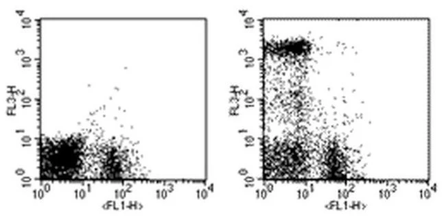
Description: The RB6-8C5 monoclonal antibody reacts with mouse Ly-6G, a 21-25 kDa protein also known as the myeloid differentiation antigen Gr-1. A GPI-linked protein, Gr-1 is expressed by the myeloid lineage in a developmentally regulated manner in the bone marrow. While monocytes only express Gr-1 transiently during their bone marrow development, the expression of Gr-1 on bone marrow granulocytes as well as on peripheral neutrophils is a good marker for these populations.
eBioscience testing indicates that in the bone marrow and lysed whole blood, the antibody clone RB6-8C5 also stains cells that express the highest levels of Ly6c (as defined by staining with antibody clone HK1.4). It is recommended that 1A8-Ly6G (Product # 9668) be used when looking at Ly-6G specific targets.
Applications Reported: The RB6-8C5 antibody has been reported for use in flow cytometric analysis.
Applications Tested: The RB6-8C5 antibody has been tested by flow cytometric analysis of mouse bone marrow cells and splenocytes. This can be used at less than or equal to 0.125 µg per test. A test is defined as the amount (µg) of antibody that will stain a cell sample in a final volume of 100 µL. Cell number should be determined empirically but can range from 10^5 to 10^8 cells/test. It is recommended that the antibody be carefully titrated for optimal performance in the assay of interest.
Light sensitivity: This tandem dye is sensitive photo-induced oxidation. Please protect this vial and stained samples from light.
Fixation: Samples can be stored in IC Fixation Buffer (Product # 00-8222) (100 µL cell sample + 100 µL IC Fixation Buffer) or 1-step Fix/Lyse Solution (Product # 00-5333) for up to 3 days in the dark at 4°C with minimal impact on brightness and FRET efficiency/compensation. Some generalizations regarding fluorophore performance after fixation can be made, but clone specific performance should be determined empirically.
Excitation: 488-561 nm; Emission: 667 nm; Laser: Blue Laser, Green Laser, Yellow-Green Laser.
Filtration: 0.2 µm post-manufacturing filtered.
For Research Use Only. Not for use in diagnostic procedures. Not for resale without express authorization.
Original: $334.00
-70%$334.00
$100.20Ly-6G/Ly-6C Monoclonal Antibody (RB6-8C5), PE-Cyanine5, eBioscience
PRODUCT DETAILS
Host: Rat
Isotype: IgG2b, kappa
Clonality: Monoclonal
Clone: RB6-8C5
Format: PE-Cyanine5
Reactivity: Ms
Application: Flow Cytometry
Tested Dilution: 0.125 µg/test
Concentration: 0.2 mg/mL
Storage: 4°C, store in dark, DO NOT FREEZE!
Formulation: PBS with 0.09% sodium azide; pH 7.2
Purification: Affinity chromatography
Data Sheet: TDS
Specific Information
Description: The RB6-8C5 monoclonal antibody reacts with mouse Ly-6G, a 21-25 kDa protein also known as the myeloid differentiation antigen Gr-1. A GPI-linked protein, Gr-1 is expressed by the myeloid lineage in a developmentally regulated manner in the bone marrow. While monocytes only express Gr-1 transiently during their bone marrow development, the expression of Gr-1 on bone marrow granulocytes as well as on peripheral neutrophils is a good marker for these populations.
eBioscience testing indicates that in the bone marrow and lysed whole blood, the antibody clone RB6-8C5 also stains cells that express the highest levels of Ly6c (as defined by staining with antibody clone HK1.4). It is recommended that 1A8-Ly6G (Product # 9668) be used when looking at Ly-6G specific targets.
Applications Reported: The RB6-8C5 antibody has been reported for use in flow cytometric analysis.
Applications Tested: The RB6-8C5 antibody has been tested by flow cytometric analysis of mouse bone marrow cells and splenocytes. This can be used at less than or equal to 0.125 µg per test. A test is defined as the amount (µg) of antibody that will stain a cell sample in a final volume of 100 µL. Cell number should be determined empirically but can range from 10^5 to 10^8 cells/test. It is recommended that the antibody be carefully titrated for optimal performance in the assay of interest.
Light sensitivity: This tandem dye is sensitive photo-induced oxidation. Please protect this vial and stained samples from light.
Fixation: Samples can be stored in IC Fixation Buffer (Product # 00-8222) (100 µL cell sample + 100 µL IC Fixation Buffer) or 1-step Fix/Lyse Solution (Product # 00-5333) for up to 3 days in the dark at 4°C with minimal impact on brightness and FRET efficiency/compensation. Some generalizations regarding fluorophore performance after fixation can be made, but clone specific performance should be determined empirically.
Excitation: 488-561 nm; Emission: 667 nm; Laser: Blue Laser, Green Laser, Yellow-Green Laser.
Filtration: 0.2 µm post-manufacturing filtered.
For Research Use Only. Not for use in diagnostic procedures. Not for resale without express authorization.
Product Information
Product Information
Shipping & Returns
Shipping & Returns
Description
PRODUCT DETAILS
Host: Rat
Isotype: IgG2b, kappa
Clonality: Monoclonal
Clone: RB6-8C5
Format: PE-Cyanine5
Reactivity: Ms
Application: Flow Cytometry
Tested Dilution: 0.125 µg/test
Concentration: 0.2 mg/mL
Storage: 4°C, store in dark, DO NOT FREEZE!
Formulation: PBS with 0.09% sodium azide; pH 7.2
Purification: Affinity chromatography
Data Sheet: TDS
Specific Information
Description: The RB6-8C5 monoclonal antibody reacts with mouse Ly-6G, a 21-25 kDa protein also known as the myeloid differentiation antigen Gr-1. A GPI-linked protein, Gr-1 is expressed by the myeloid lineage in a developmentally regulated manner in the bone marrow. While monocytes only express Gr-1 transiently during their bone marrow development, the expression of Gr-1 on bone marrow granulocytes as well as on peripheral neutrophils is a good marker for these populations.
eBioscience testing indicates that in the bone marrow and lysed whole blood, the antibody clone RB6-8C5 also stains cells that express the highest levels of Ly6c (as defined by staining with antibody clone HK1.4). It is recommended that 1A8-Ly6G (Product # 9668) be used when looking at Ly-6G specific targets.
Applications Reported: The RB6-8C5 antibody has been reported for use in flow cytometric analysis.
Applications Tested: The RB6-8C5 antibody has been tested by flow cytometric analysis of mouse bone marrow cells and splenocytes. This can be used at less than or equal to 0.125 µg per test. A test is defined as the amount (µg) of antibody that will stain a cell sample in a final volume of 100 µL. Cell number should be determined empirically but can range from 10^5 to 10^8 cells/test. It is recommended that the antibody be carefully titrated for optimal performance in the assay of interest.
Light sensitivity: This tandem dye is sensitive photo-induced oxidation. Please protect this vial and stained samples from light.
Fixation: Samples can be stored in IC Fixation Buffer (Product # 00-8222) (100 µL cell sample + 100 µL IC Fixation Buffer) or 1-step Fix/Lyse Solution (Product # 00-5333) for up to 3 days in the dark at 4°C with minimal impact on brightness and FRET efficiency/compensation. Some generalizations regarding fluorophore performance after fixation can be made, but clone specific performance should be determined empirically.
Excitation: 488-561 nm; Emission: 667 nm; Laser: Blue Laser, Green Laser, Yellow-Green Laser.
Filtration: 0.2 µm post-manufacturing filtered.
For Research Use Only. Not for use in diagnostic procedures. Not for resale without express authorization.


