LAP (Latency Associated peptide) Monoclonal Antibody (TW7-16B4), PE-Cyanine7, eBioscience
PRODUCT DETAILS
Host: Mouse
Isotype: IgG1, kappa
Clonality: Monoclonal
Clone: TW7-16B4
Format: PE-Cyanine7
Reactivity: Ms
Application: Flow Cytometry
Tested Dilution: 0.125 µg/test
Concentration: 0.2 mg/mL
Storage: 4°C, store in dark, DO NOT FREEZE!
Formulation: PBS with 0.09% sodium azide; pH 7.2
Purification: Affinity chromatography
Data Sheet: TDS
Specific Information
Description: The TW7-16B4 monoclonal antibody reacts with mouse latency associated peptide (LAP, pro-TGF beta 1, LAP/TGF beta 1). Many different cells produce TGF beta and it mediates effects on the proliferation, differentiation and function of many cell types. TGF beta is synthesized as a precursor that contains LAP at the N-terminus and mature TGF beta at the C-terminus. Processing and cleavage of the precursor protein between amino acids 278 and 279 results in the formation of LAP dimers and TGF beta dimers that then non-covalently associate with each other to form the small latent TGF beta complex. LAP is secreted and can be found in the extracellular matrix. In addition, LAP can also be expressed on platelets and activated regulatory T cells. It is believed that this surface-expressed LAP is due to the binding of LAP to GARP (LRRC32), which is a transmembrane protein that is also found at high levels on platelets and activated regulatory T cells.
Applications Reported: This TW7-16B4 antibody has been reported for use in flow cytometric analysis.
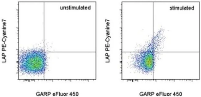
Applications Tested: This TW7-16B4 antibody has been tested by flow cytometric analysis of stimulated mouse splenocytes. This can be used at less than or equal to 0.125 µg per test. A test is defined as the amount (µg) of antibody that will stain a cell sample in a final volume of 100 µL. Cell number should be determined empirically but can range from 10^5 to 10^8 cells/test. It is recommended that the antibody be carefully titrated for optimal performance in the assay of interest.
Light sensitivity: This tandem dye is sensitive photo-induced oxidation. Please protect this vial and stained samples from light.
Fixation: Samples can be stored in IC Fixation Buffer (Product # 00-822-49) (100 µL cell sample + 100 µL IC Fixation Buffer) or 1-step Fix/Lyse Solution (Product # 00-5333-54) for up to 3 days in the dark at 4°C with minimal impact on brightness and FRET efficiency/compensation. Some generalizations regarding fluorophore performance after fixation can be made, but clone specific performance should be determined empirically.
Excitation: 488-561 nm; Emission: 775 nm; Laser: Blue Laser, Green Laser, Yellow-Green Laser.
Filtration: 0.2 µm post-manufacturing filtered.
For Research Use Only. Not for use in diagnostic procedures. Not for resale without express authorization.
Original: $405.00
-70%$405.00
$121.50LAP (Latency Associated peptide) Monoclonal Antibody (TW7-16B4), PE-Cyanine7, eBioscience
PRODUCT DETAILS
Host: Mouse
Isotype: IgG1, kappa
Clonality: Monoclonal
Clone: TW7-16B4
Format: PE-Cyanine7
Reactivity: Ms
Application: Flow Cytometry
Tested Dilution: 0.125 µg/test
Concentration: 0.2 mg/mL
Storage: 4°C, store in dark, DO NOT FREEZE!
Formulation: PBS with 0.09% sodium azide; pH 7.2
Purification: Affinity chromatography
Data Sheet: TDS
Specific Information
Description: The TW7-16B4 monoclonal antibody reacts with mouse latency associated peptide (LAP, pro-TGF beta 1, LAP/TGF beta 1). Many different cells produce TGF beta and it mediates effects on the proliferation, differentiation and function of many cell types. TGF beta is synthesized as a precursor that contains LAP at the N-terminus and mature TGF beta at the C-terminus. Processing and cleavage of the precursor protein between amino acids 278 and 279 results in the formation of LAP dimers and TGF beta dimers that then non-covalently associate with each other to form the small latent TGF beta complex. LAP is secreted and can be found in the extracellular matrix. In addition, LAP can also be expressed on platelets and activated regulatory T cells. It is believed that this surface-expressed LAP is due to the binding of LAP to GARP (LRRC32), which is a transmembrane protein that is also found at high levels on platelets and activated regulatory T cells.
Applications Reported: This TW7-16B4 antibody has been reported for use in flow cytometric analysis.
Applications Tested: This TW7-16B4 antibody has been tested by flow cytometric analysis of stimulated mouse splenocytes. This can be used at less than or equal to 0.125 µg per test. A test is defined as the amount (µg) of antibody that will stain a cell sample in a final volume of 100 µL. Cell number should be determined empirically but can range from 10^5 to 10^8 cells/test. It is recommended that the antibody be carefully titrated for optimal performance in the assay of interest.
Light sensitivity: This tandem dye is sensitive photo-induced oxidation. Please protect this vial and stained samples from light.
Fixation: Samples can be stored in IC Fixation Buffer (Product # 00-822-49) (100 µL cell sample + 100 µL IC Fixation Buffer) or 1-step Fix/Lyse Solution (Product # 00-5333-54) for up to 3 days in the dark at 4°C with minimal impact on brightness and FRET efficiency/compensation. Some generalizations regarding fluorophore performance after fixation can be made, but clone specific performance should be determined empirically.
Excitation: 488-561 nm; Emission: 775 nm; Laser: Blue Laser, Green Laser, Yellow-Green Laser.
Filtration: 0.2 µm post-manufacturing filtered.
For Research Use Only. Not for use in diagnostic procedures. Not for resale without express authorization.
Product Information
Product Information
Shipping & Returns
Shipping & Returns
Description
PRODUCT DETAILS
Host: Mouse
Isotype: IgG1, kappa
Clonality: Monoclonal
Clone: TW7-16B4
Format: PE-Cyanine7
Reactivity: Ms
Application: Flow Cytometry
Tested Dilution: 0.125 µg/test
Concentration: 0.2 mg/mL
Storage: 4°C, store in dark, DO NOT FREEZE!
Formulation: PBS with 0.09% sodium azide; pH 7.2
Purification: Affinity chromatography
Data Sheet: TDS
Specific Information
Description: The TW7-16B4 monoclonal antibody reacts with mouse latency associated peptide (LAP, pro-TGF beta 1, LAP/TGF beta 1). Many different cells produce TGF beta and it mediates effects on the proliferation, differentiation and function of many cell types. TGF beta is synthesized as a precursor that contains LAP at the N-terminus and mature TGF beta at the C-terminus. Processing and cleavage of the precursor protein between amino acids 278 and 279 results in the formation of LAP dimers and TGF beta dimers that then non-covalently associate with each other to form the small latent TGF beta complex. LAP is secreted and can be found in the extracellular matrix. In addition, LAP can also be expressed on platelets and activated regulatory T cells. It is believed that this surface-expressed LAP is due to the binding of LAP to GARP (LRRC32), which is a transmembrane protein that is also found at high levels on platelets and activated regulatory T cells.
Applications Reported: This TW7-16B4 antibody has been reported for use in flow cytometric analysis.
Applications Tested: This TW7-16B4 antibody has been tested by flow cytometric analysis of stimulated mouse splenocytes. This can be used at less than or equal to 0.125 µg per test. A test is defined as the amount (µg) of antibody that will stain a cell sample in a final volume of 100 µL. Cell number should be determined empirically but can range from 10^5 to 10^8 cells/test. It is recommended that the antibody be carefully titrated for optimal performance in the assay of interest.
Light sensitivity: This tandem dye is sensitive photo-induced oxidation. Please protect this vial and stained samples from light.
Fixation: Samples can be stored in IC Fixation Buffer (Product # 00-822-49) (100 µL cell sample + 100 µL IC Fixation Buffer) or 1-step Fix/Lyse Solution (Product # 00-5333-54) for up to 3 days in the dark at 4°C with minimal impact on brightness and FRET efficiency/compensation. Some generalizations regarding fluorophore performance after fixation can be made, but clone specific performance should be determined empirically.
Excitation: 488-561 nm; Emission: 775 nm; Laser: Blue Laser, Green Laser, Yellow-Green Laser.
Filtration: 0.2 µm post-manufacturing filtered.
For Research Use Only. Not for use in diagnostic procedures. Not for resale without express authorization.


