IRF7 Monoclonal Antibody (RDP4ND4), Alexa Fluor 647, eBioscience
PRODUCT DETAILS
Host: Rat
Isotype: IgG2a, kappa
Clonality: Monoclonal
Clone: RDP4ND4
Format: Alexa Fluor 647
Reactivity: Hu
Application: Flow Cytometry
Tested Dilution: 5 µL (0.06 µg)/test
Concentration: 5 μL/Test
Storage: 4°C, store in dark, DO NOT FREEZE!
Formulation: PBS with BSA and 0.09% sodium azide; pH 7.2
Purification: Affinity chromatography
Data Sheet: TDS
Specific Information
Description: The monoclonal antibody RDP4ND4 recognizes human interferon regulatory factor 7 (IRF7), a 54 kDa member of the interferon regulatory transcription factor family. It is essential for the antiviral innate immune response and the activation of type I IFN genes. IRF7 is constitutively expressed in the cytoplasm of plasmacytoid dendritic cells, B cells and monocytes, but it is strongly induced in response to virus infection and interferon stimulation.
IRF7 can activate IFN-beta and IFN-alpha genes via the virus-activated, MyD88-independent pathway, or the TLR-activated, MyD88-dependent pathway. Virus exposure induces IRF7, through toll-like receptors TLR7, TLR8, and TLR9, to promote IFN gene expression and disrupt infection. IRF7 exists in an inactive form until phosphorylated and its phosphorylation leads to dimerization and nuclear translocation of IRF7 where it aids in activating target genes. The IRF7 gene is upregulated by interferon stimulation through the Jak-STAT pathway in a positive feedback loop.
Applications Reported: This RDP4ND4 antibody has been reported for use in flow cytometric analysis.
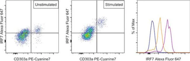
Applications Tested: This RDP4ND4 antibody has been pre-diluted and tested by flow cytometric analysis of stimulated normal human peripheral blood cells using the Foxp3/Transcription Factor Staining Buffer Set (Product # 00-5523-00) and protocol. Please refer to BestProtocols®: Protocol B: One step protocol for (nuclear) intracellular proteins located under the Resources Tab online. This may be used at 5 µL (0.06 µg) per test. A test is defined as the amount (µg) of antibody that will stain a cell sample in a final volume of 100 µL. Cell number should be determined empirically but can range from 10^5 to 10^8 cells/test.
Excitation: 633-647 nm; Emission: 668 nm; Laser: Red Laser.
For Research Use Only. Not for use in diagnostic procedures. Not for resale without express authorization.
Original: $506.00
-70%$506.00
$151.80IRF7 Monoclonal Antibody (RDP4ND4), Alexa Fluor 647, eBioscience
PRODUCT DETAILS
Host: Rat
Isotype: IgG2a, kappa
Clonality: Monoclonal
Clone: RDP4ND4
Format: Alexa Fluor 647
Reactivity: Hu
Application: Flow Cytometry
Tested Dilution: 5 µL (0.06 µg)/test
Concentration: 5 μL/Test
Storage: 4°C, store in dark, DO NOT FREEZE!
Formulation: PBS with BSA and 0.09% sodium azide; pH 7.2
Purification: Affinity chromatography
Data Sheet: TDS
Specific Information
Description: The monoclonal antibody RDP4ND4 recognizes human interferon regulatory factor 7 (IRF7), a 54 kDa member of the interferon regulatory transcription factor family. It is essential for the antiviral innate immune response and the activation of type I IFN genes. IRF7 is constitutively expressed in the cytoplasm of plasmacytoid dendritic cells, B cells and monocytes, but it is strongly induced in response to virus infection and interferon stimulation.
IRF7 can activate IFN-beta and IFN-alpha genes via the virus-activated, MyD88-independent pathway, or the TLR-activated, MyD88-dependent pathway. Virus exposure induces IRF7, through toll-like receptors TLR7, TLR8, and TLR9, to promote IFN gene expression and disrupt infection. IRF7 exists in an inactive form until phosphorylated and its phosphorylation leads to dimerization and nuclear translocation of IRF7 where it aids in activating target genes. The IRF7 gene is upregulated by interferon stimulation through the Jak-STAT pathway in a positive feedback loop.
Applications Reported: This RDP4ND4 antibody has been reported for use in flow cytometric analysis.
Applications Tested: This RDP4ND4 antibody has been pre-diluted and tested by flow cytometric analysis of stimulated normal human peripheral blood cells using the Foxp3/Transcription Factor Staining Buffer Set (Product # 00-5523-00) and protocol. Please refer to BestProtocols®: Protocol B: One step protocol for (nuclear) intracellular proteins located under the Resources Tab online. This may be used at 5 µL (0.06 µg) per test. A test is defined as the amount (µg) of antibody that will stain a cell sample in a final volume of 100 µL. Cell number should be determined empirically but can range from 10^5 to 10^8 cells/test.
Excitation: 633-647 nm; Emission: 668 nm; Laser: Red Laser.
For Research Use Only. Not for use in diagnostic procedures. Not for resale without express authorization.
Product Information
Product Information
Shipping & Returns
Shipping & Returns
Description
PRODUCT DETAILS
Host: Rat
Isotype: IgG2a, kappa
Clonality: Monoclonal
Clone: RDP4ND4
Format: Alexa Fluor 647
Reactivity: Hu
Application: Flow Cytometry
Tested Dilution: 5 µL (0.06 µg)/test
Concentration: 5 μL/Test
Storage: 4°C, store in dark, DO NOT FREEZE!
Formulation: PBS with BSA and 0.09% sodium azide; pH 7.2
Purification: Affinity chromatography
Data Sheet: TDS
Specific Information
Description: The monoclonal antibody RDP4ND4 recognizes human interferon regulatory factor 7 (IRF7), a 54 kDa member of the interferon regulatory transcription factor family. It is essential for the antiviral innate immune response and the activation of type I IFN genes. IRF7 is constitutively expressed in the cytoplasm of plasmacytoid dendritic cells, B cells and monocytes, but it is strongly induced in response to virus infection and interferon stimulation.
IRF7 can activate IFN-beta and IFN-alpha genes via the virus-activated, MyD88-independent pathway, or the TLR-activated, MyD88-dependent pathway. Virus exposure induces IRF7, through toll-like receptors TLR7, TLR8, and TLR9, to promote IFN gene expression and disrupt infection. IRF7 exists in an inactive form until phosphorylated and its phosphorylation leads to dimerization and nuclear translocation of IRF7 where it aids in activating target genes. The IRF7 gene is upregulated by interferon stimulation through the Jak-STAT pathway in a positive feedback loop.
Applications Reported: This RDP4ND4 antibody has been reported for use in flow cytometric analysis.
Applications Tested: This RDP4ND4 antibody has been pre-diluted and tested by flow cytometric analysis of stimulated normal human peripheral blood cells using the Foxp3/Transcription Factor Staining Buffer Set (Product # 00-5523-00) and protocol. Please refer to BestProtocols®: Protocol B: One step protocol for (nuclear) intracellular proteins located under the Resources Tab online. This may be used at 5 µL (0.06 µg) per test. A test is defined as the amount (µg) of antibody that will stain a cell sample in a final volume of 100 µL. Cell number should be determined empirically but can range from 10^5 to 10^8 cells/test.
Excitation: 633-647 nm; Emission: 668 nm; Laser: Red Laser.
For Research Use Only. Not for use in diagnostic procedures. Not for resale without express authorization.


