IL-27 p28 Monoclonal Antibody (3D1p28), eFluor 660, eBioscience
PRODUCT DETAILS
Host: Mouse
Isotype: IgG1, kappa
Clonality: Monoclonal
Clone: 3D1p28
Format: eFluor 660
Reactivity: Hu
Application: Flow Cytometry
Tested Dilution: 5 µL (0.03 µg)/test
Concentration: 5 μL/Test
Storage: 4°C, store in dark, DO NOT FREEZE!
Formulation: PBS with BSA and 0.09% sodium azide; pH 7.2
Purification: Affinity chromatography
Data Sheet: TDS
Specific Information
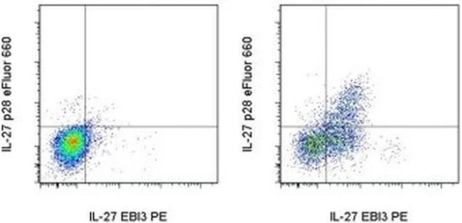
Description: This 3D1p28 monoclonal antibody reacts with the human p28 subunit of IL-27. Interleukin-27 (IL-27) is a member of the IL-12 family, a subgroup of the IL-6 family of cytokines. It is a heterodimer of the subunits EBI3 (Epstein-Barr Virus Induced Gene 3), which is homologous to the p40 subunit shared by IL-12 and IL-23, and p28 (IL-30), which is homologous to p35. IL-27 is produced by activated dendritic cells and macrophages in response to TLR ligands and inflammatory cytokines. IL-27 has been shown to have both pro-inflammatory and anti-inflammatory effects. It influences the commitment of CD4+ T-cells toward the Th1 lineage by inducing the expression of the transcription factor T-bet and the upregulation of IL-12R beta2. Its anti-inflammatory functions include the suppression of Th2 and Th17 proliferation and differentiation.
The IL-27 receptor shares one subunit, gp130, with other members of the IL-6 family. The subunit WSX-1 (IL-27R alpha, TCCR) is unique to IL-27 and is believed to be the only part of the receptor that interacts with the cytokine. IL-27R activation leads to the phosphorylation of Jak/STAT proteins, with STAT1 and STAT3 being critical to the function of IL-27.
Recent evidence suggests that the p28 subunit may also be secreted independently of EBI3 and have functions distinct from the IL-27 heterodimer: it is believed to not only antagonize the activity of IL-27, but also inhibit signaling of other gp130 ligands, such as IL-6 and IL-11.
Applications Reported: This 3D1p28 antibody has been reported for use in intracellular staining followed by flow cytometric analysis.
Applications Tested: This 3D1p28 antibody has been pre-titrated and tested by intracellular staining followed by flow cytometric analysis. This can be used at 5 µL (0.03 µg) per test. A test is defined as the amount (µg) of antibody that will stain a cell sample in a final volume of 100 µL. Cell number should be determined empirically but can range from 10^5 to 10^8 cells/test.
eFluor® 660 is a replacement for Alexa Fluor® 647. eFluor® 660 emits at 659 nm and is excited with the red laser (633 nm). Please make sure that your instrument is capable of detecting this fluorochome.
Excitation: 633-647 nm; Emission: 668 nm; Laser: Red Laser.
Filtration: 0.2 µm post-manufacturing filtered.
For Research Use Only. Not for use in diagnostic procedures. Not for resale without express authorization.
Original: $418.00
-70%$418.00
$125.40IL-27 p28 Monoclonal Antibody (3D1p28), eFluor 660, eBioscience
PRODUCT DETAILS
Host: Mouse
Isotype: IgG1, kappa
Clonality: Monoclonal
Clone: 3D1p28
Format: eFluor 660
Reactivity: Hu
Application: Flow Cytometry
Tested Dilution: 5 µL (0.03 µg)/test
Concentration: 5 μL/Test
Storage: 4°C, store in dark, DO NOT FREEZE!
Formulation: PBS with BSA and 0.09% sodium azide; pH 7.2
Purification: Affinity chromatography
Data Sheet: TDS
Specific Information
Description: This 3D1p28 monoclonal antibody reacts with the human p28 subunit of IL-27. Interleukin-27 (IL-27) is a member of the IL-12 family, a subgroup of the IL-6 family of cytokines. It is a heterodimer of the subunits EBI3 (Epstein-Barr Virus Induced Gene 3), which is homologous to the p40 subunit shared by IL-12 and IL-23, and p28 (IL-30), which is homologous to p35. IL-27 is produced by activated dendritic cells and macrophages in response to TLR ligands and inflammatory cytokines. IL-27 has been shown to have both pro-inflammatory and anti-inflammatory effects. It influences the commitment of CD4+ T-cells toward the Th1 lineage by inducing the expression of the transcription factor T-bet and the upregulation of IL-12R beta2. Its anti-inflammatory functions include the suppression of Th2 and Th17 proliferation and differentiation.
The IL-27 receptor shares one subunit, gp130, with other members of the IL-6 family. The subunit WSX-1 (IL-27R alpha, TCCR) is unique to IL-27 and is believed to be the only part of the receptor that interacts with the cytokine. IL-27R activation leads to the phosphorylation of Jak/STAT proteins, with STAT1 and STAT3 being critical to the function of IL-27.
Recent evidence suggests that the p28 subunit may also be secreted independently of EBI3 and have functions distinct from the IL-27 heterodimer: it is believed to not only antagonize the activity of IL-27, but also inhibit signaling of other gp130 ligands, such as IL-6 and IL-11.
Applications Reported: This 3D1p28 antibody has been reported for use in intracellular staining followed by flow cytometric analysis.
Applications Tested: This 3D1p28 antibody has been pre-titrated and tested by intracellular staining followed by flow cytometric analysis. This can be used at 5 µL (0.03 µg) per test. A test is defined as the amount (µg) of antibody that will stain a cell sample in a final volume of 100 µL. Cell number should be determined empirically but can range from 10^5 to 10^8 cells/test.
eFluor® 660 is a replacement for Alexa Fluor® 647. eFluor® 660 emits at 659 nm and is excited with the red laser (633 nm). Please make sure that your instrument is capable of detecting this fluorochome.
Excitation: 633-647 nm; Emission: 668 nm; Laser: Red Laser.
Filtration: 0.2 µm post-manufacturing filtered.
For Research Use Only. Not for use in diagnostic procedures. Not for resale without express authorization.
Product Information
Product Information
Shipping & Returns
Shipping & Returns
Description
PRODUCT DETAILS
Host: Mouse
Isotype: IgG1, kappa
Clonality: Monoclonal
Clone: 3D1p28
Format: eFluor 660
Reactivity: Hu
Application: Flow Cytometry
Tested Dilution: 5 µL (0.03 µg)/test
Concentration: 5 μL/Test
Storage: 4°C, store in dark, DO NOT FREEZE!
Formulation: PBS with BSA and 0.09% sodium azide; pH 7.2
Purification: Affinity chromatography
Data Sheet: TDS
Specific Information
Description: This 3D1p28 monoclonal antibody reacts with the human p28 subunit of IL-27. Interleukin-27 (IL-27) is a member of the IL-12 family, a subgroup of the IL-6 family of cytokines. It is a heterodimer of the subunits EBI3 (Epstein-Barr Virus Induced Gene 3), which is homologous to the p40 subunit shared by IL-12 and IL-23, and p28 (IL-30), which is homologous to p35. IL-27 is produced by activated dendritic cells and macrophages in response to TLR ligands and inflammatory cytokines. IL-27 has been shown to have both pro-inflammatory and anti-inflammatory effects. It influences the commitment of CD4+ T-cells toward the Th1 lineage by inducing the expression of the transcription factor T-bet and the upregulation of IL-12R beta2. Its anti-inflammatory functions include the suppression of Th2 and Th17 proliferation and differentiation.
The IL-27 receptor shares one subunit, gp130, with other members of the IL-6 family. The subunit WSX-1 (IL-27R alpha, TCCR) is unique to IL-27 and is believed to be the only part of the receptor that interacts with the cytokine. IL-27R activation leads to the phosphorylation of Jak/STAT proteins, with STAT1 and STAT3 being critical to the function of IL-27.
Recent evidence suggests that the p28 subunit may also be secreted independently of EBI3 and have functions distinct from the IL-27 heterodimer: it is believed to not only antagonize the activity of IL-27, but also inhibit signaling of other gp130 ligands, such as IL-6 and IL-11.
Applications Reported: This 3D1p28 antibody has been reported for use in intracellular staining followed by flow cytometric analysis.
Applications Tested: This 3D1p28 antibody has been pre-titrated and tested by intracellular staining followed by flow cytometric analysis. This can be used at 5 µL (0.03 µg) per test. A test is defined as the amount (µg) of antibody that will stain a cell sample in a final volume of 100 µL. Cell number should be determined empirically but can range from 10^5 to 10^8 cells/test.
eFluor® 660 is a replacement for Alexa Fluor® 647. eFluor® 660 emits at 659 nm and is excited with the red laser (633 nm). Please make sure that your instrument is capable of detecting this fluorochome.
Excitation: 633-647 nm; Emission: 668 nm; Laser: Red Laser.
Filtration: 0.2 µm post-manufacturing filtered.
For Research Use Only. Not for use in diagnostic procedures. Not for resale without express authorization.


