HIF-1 alpha Monoclonal Antibody (Mgc3), PE, eBioscience
PRODUCT DETAILS
Host: Mouse
Isotype: IgG1, kappa
Clonality: Monoclonal
Clone: Mgc3
Format: PE
Reactivity: Hu, Ms
Application: Flow Cytometry
Tested Dilution: 1.0 µg/test
Concentration: 0.2 mg/mL
Storage: 4°C, store in dark, DO NOT FREEZE!
Formulation: PBS with 0.09% sodium azide; pH 7.2
Purification: Affinity chromatography
Data Sheet: TDS
Specific Information
Description: This Mgc3 monoclonal antibody detects hypoxia-inducible factor 1 alpha (HIF-1 alpha) from human, non-human primate, bovine, mouse and porcine cells. This antibody does not cross-react with ARNT or the related HIF-2 alpha.
Mgc3 has been successfully used in western blotting, immunofluorescence, immunocytochemistry, immunoprecipitation, flow cytometry and gel shift procedures. By western blot, this antibody detects a ~116 kDa protein representing HIF-1 alpha after hypoxic induction in COS cells. Immunofluorescence staining of HIF-1 alpha in COS-7 cells with clone Mgc3 yields nuclear staining after exposing cells to 1% oxygen for 4 hours. In gel shift assay experiments, the Mgc3 antibody detected only mouse and not human HIF-1 alpha.
Applications Reported: This Mgc3 antibody has been reported for use in flow cytometric analysis.
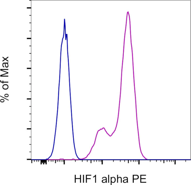
Applications Tested: This Mgc3 antibody has been tested by flow cytometric analysis of DFOA stimulated HeLa cells using the methanol fixation protocol. This may be used at less than or equal to 1.0 µg/mL per test. A test is defined as the amount (µg) of antibody that will stain a cell sample in a final volume of 100 µL. Cell number should be determined empirically but can range from 10^5 to 10^8 cells/test. It is recommended that the antibody be carefully titrated for optimal performance in the assay of interest.
Excitation: 488-561 nm; Emission: 578 nm; Laser: Blue Laser, Green Laser, Yellow-Green Laser
For Research Use Only. Not for use in diagnostic procedures. Not for resale without express authorization.
Original: $379.00
-70%$379.00
$113.70HIF-1 alpha Monoclonal Antibody (Mgc3), PE, eBioscience
PRODUCT DETAILS
Host: Mouse
Isotype: IgG1, kappa
Clonality: Monoclonal
Clone: Mgc3
Format: PE
Reactivity: Hu, Ms
Application: Flow Cytometry
Tested Dilution: 1.0 µg/test
Concentration: 0.2 mg/mL
Storage: 4°C, store in dark, DO NOT FREEZE!
Formulation: PBS with 0.09% sodium azide; pH 7.2
Purification: Affinity chromatography
Data Sheet: TDS
Specific Information
Description: This Mgc3 monoclonal antibody detects hypoxia-inducible factor 1 alpha (HIF-1 alpha) from human, non-human primate, bovine, mouse and porcine cells. This antibody does not cross-react with ARNT or the related HIF-2 alpha.
Mgc3 has been successfully used in western blotting, immunofluorescence, immunocytochemistry, immunoprecipitation, flow cytometry and gel shift procedures. By western blot, this antibody detects a ~116 kDa protein representing HIF-1 alpha after hypoxic induction in COS cells. Immunofluorescence staining of HIF-1 alpha in COS-7 cells with clone Mgc3 yields nuclear staining after exposing cells to 1% oxygen for 4 hours. In gel shift assay experiments, the Mgc3 antibody detected only mouse and not human HIF-1 alpha.
Applications Reported: This Mgc3 antibody has been reported for use in flow cytometric analysis.
Applications Tested: This Mgc3 antibody has been tested by flow cytometric analysis of DFOA stimulated HeLa cells using the methanol fixation protocol. This may be used at less than or equal to 1.0 µg/mL per test. A test is defined as the amount (µg) of antibody that will stain a cell sample in a final volume of 100 µL. Cell number should be determined empirically but can range from 10^5 to 10^8 cells/test. It is recommended that the antibody be carefully titrated for optimal performance in the assay of interest.
Excitation: 488-561 nm; Emission: 578 nm; Laser: Blue Laser, Green Laser, Yellow-Green Laser
For Research Use Only. Not for use in diagnostic procedures. Not for resale without express authorization.
Product Information
Product Information
Shipping & Returns
Shipping & Returns
Description
PRODUCT DETAILS
Host: Mouse
Isotype: IgG1, kappa
Clonality: Monoclonal
Clone: Mgc3
Format: PE
Reactivity: Hu, Ms
Application: Flow Cytometry
Tested Dilution: 1.0 µg/test
Concentration: 0.2 mg/mL
Storage: 4°C, store in dark, DO NOT FREEZE!
Formulation: PBS with 0.09% sodium azide; pH 7.2
Purification: Affinity chromatography
Data Sheet: TDS
Specific Information
Description: This Mgc3 monoclonal antibody detects hypoxia-inducible factor 1 alpha (HIF-1 alpha) from human, non-human primate, bovine, mouse and porcine cells. This antibody does not cross-react with ARNT or the related HIF-2 alpha.
Mgc3 has been successfully used in western blotting, immunofluorescence, immunocytochemistry, immunoprecipitation, flow cytometry and gel shift procedures. By western blot, this antibody detects a ~116 kDa protein representing HIF-1 alpha after hypoxic induction in COS cells. Immunofluorescence staining of HIF-1 alpha in COS-7 cells with clone Mgc3 yields nuclear staining after exposing cells to 1% oxygen for 4 hours. In gel shift assay experiments, the Mgc3 antibody detected only mouse and not human HIF-1 alpha.
Applications Reported: This Mgc3 antibody has been reported for use in flow cytometric analysis.
Applications Tested: This Mgc3 antibody has been tested by flow cytometric analysis of DFOA stimulated HeLa cells using the methanol fixation protocol. This may be used at less than or equal to 1.0 µg/mL per test. A test is defined as the amount (µg) of antibody that will stain a cell sample in a final volume of 100 µL. Cell number should be determined empirically but can range from 10^5 to 10^8 cells/test. It is recommended that the antibody be carefully titrated for optimal performance in the assay of interest.
Excitation: 488-561 nm; Emission: 578 nm; Laser: Blue Laser, Green Laser, Yellow-Green Laser
For Research Use Only. Not for use in diagnostic procedures. Not for resale without express authorization.


