Granzyme A Monoclonal Antibody (CB9), PE-Cyanine7, eBioscience
PRODUCT DETAILS
Host: Mouse
Isotype: IgG1, kappa
Clonality: Monoclonal
Clone: CB9
Format: PE-Cyanine7
Reactivity: Hu
Application: Flow Cytometry
Tested Dilution: 5 µL (0.25 µg)/test
Concentration: 5 μL/Test
Storage: 4°C, store in dark, DO NOT FREEZE!
Formulation: PBS with BSA and 0.09% sodium azide; pH 7.2
Purification: Affinity chromatography
Data Sheet: TDS
Specific Information
Description: This CB9 monoclonal antibody reacts with human Granzyme A. Granzyme A is the most abundantly expressed of the granzyme serine proteases, which are proteins released from the granules of NK cells and cytotoxic T lymphocytes that induce death in target cells by cleavage of intracellular substrates. They play a critical role in immune defense against viruses, tumors, and intracellular bacteria. Granzyme A activates caspase-independent cell death pathways morphologically similar to apoptosis and characterized by mitochondrial and DNA damage. It may also play a role in inflammation, as the precursor form of IL-1 beta (pro-IL-1 beta) is among its target substrates. Granzyme A shares overlapping substrate specificity with the closely-related Granzyme K, which is believed to account for the minimal decrease in cytotoxicity of Granzyme A-deficient CTL.
Applications Reported: This CB9 antibody has been reported for use in intracellular staining followed by flow cytometric analysis.
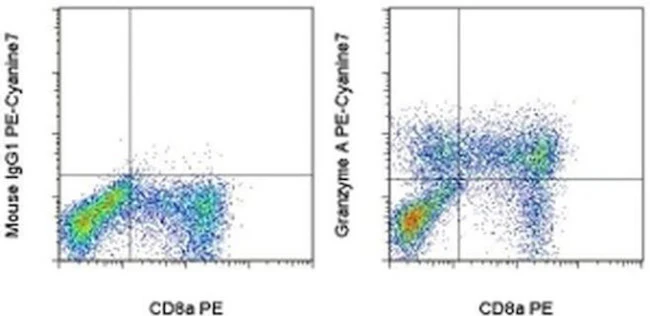
Applications Tested: This CB9 antibody has been pre-titrated and tested by intracellular staining followed by flow cytometric analysis of stimulated normal human peripheral blood cells using the Intracellular Fixation & Permeabilization Buffer Set (Product # 88-8824-00) and protocol. Please refer to BestProtocols®: Protocol A: Two step protocol for (cytoplasmic) intracellular proteins located under the Resources Tab online. This can be used at 5 µL (0.25 µg) per test. A test is defined as the amount (µg) of antibody that will stain a cell sample in a final volume of 100 µL. Cell number should be determined empirically but can range from 10^5 to 10^8 cells/test.
Light sensitivity: This tandem dye is sensitive to photo-induced oxidation. Please protect this vial and stained samples from light.
Fixation: Samples can be stored in IC Fixation Buffer (Product # 00-822-49) (100 µL of cell sample + 100 µL of IC Fixation Buffer) or 1-step Fix/Lyse Solution (Product # 00-5333-54) for up to 3 days in the dark at 4°C with minimal impact on brightness and FRET efficiency/compensation. Some generalizations regarding fluorophore performance after fixation can be made, but clone specific performance should be determined empirically.
Excitation: 488-561 nm; Emission: 775 nm; Laser: Blue Laser, Green Laser, Yellow-Green Laser.
Filtration: 0.2 µm post-manufacturing filtered.
For Research Use Only. Not for use in diagnostic procedures. Not for resale without express authorization.
Original: $500.00
-70%$500.00
$150.00Granzyme A Monoclonal Antibody (CB9), PE-Cyanine7, eBioscience
PRODUCT DETAILS
Host: Mouse
Isotype: IgG1, kappa
Clonality: Monoclonal
Clone: CB9
Format: PE-Cyanine7
Reactivity: Hu
Application: Flow Cytometry
Tested Dilution: 5 µL (0.25 µg)/test
Concentration: 5 μL/Test
Storage: 4°C, store in dark, DO NOT FREEZE!
Formulation: PBS with BSA and 0.09% sodium azide; pH 7.2
Purification: Affinity chromatography
Data Sheet: TDS
Specific Information
Description: This CB9 monoclonal antibody reacts with human Granzyme A. Granzyme A is the most abundantly expressed of the granzyme serine proteases, which are proteins released from the granules of NK cells and cytotoxic T lymphocytes that induce death in target cells by cleavage of intracellular substrates. They play a critical role in immune defense against viruses, tumors, and intracellular bacteria. Granzyme A activates caspase-independent cell death pathways morphologically similar to apoptosis and characterized by mitochondrial and DNA damage. It may also play a role in inflammation, as the precursor form of IL-1 beta (pro-IL-1 beta) is among its target substrates. Granzyme A shares overlapping substrate specificity with the closely-related Granzyme K, which is believed to account for the minimal decrease in cytotoxicity of Granzyme A-deficient CTL.
Applications Reported: This CB9 antibody has been reported for use in intracellular staining followed by flow cytometric analysis.
Applications Tested: This CB9 antibody has been pre-titrated and tested by intracellular staining followed by flow cytometric analysis of stimulated normal human peripheral blood cells using the Intracellular Fixation & Permeabilization Buffer Set (Product # 88-8824-00) and protocol. Please refer to BestProtocols®: Protocol A: Two step protocol for (cytoplasmic) intracellular proteins located under the Resources Tab online. This can be used at 5 µL (0.25 µg) per test. A test is defined as the amount (µg) of antibody that will stain a cell sample in a final volume of 100 µL. Cell number should be determined empirically but can range from 10^5 to 10^8 cells/test.
Light sensitivity: This tandem dye is sensitive to photo-induced oxidation. Please protect this vial and stained samples from light.
Fixation: Samples can be stored in IC Fixation Buffer (Product # 00-822-49) (100 µL of cell sample + 100 µL of IC Fixation Buffer) or 1-step Fix/Lyse Solution (Product # 00-5333-54) for up to 3 days in the dark at 4°C with minimal impact on brightness and FRET efficiency/compensation. Some generalizations regarding fluorophore performance after fixation can be made, but clone specific performance should be determined empirically.
Excitation: 488-561 nm; Emission: 775 nm; Laser: Blue Laser, Green Laser, Yellow-Green Laser.
Filtration: 0.2 µm post-manufacturing filtered.
For Research Use Only. Not for use in diagnostic procedures. Not for resale without express authorization.
Product Information
Product Information
Shipping & Returns
Shipping & Returns
Description
PRODUCT DETAILS
Host: Mouse
Isotype: IgG1, kappa
Clonality: Monoclonal
Clone: CB9
Format: PE-Cyanine7
Reactivity: Hu
Application: Flow Cytometry
Tested Dilution: 5 µL (0.25 µg)/test
Concentration: 5 μL/Test
Storage: 4°C, store in dark, DO NOT FREEZE!
Formulation: PBS with BSA and 0.09% sodium azide; pH 7.2
Purification: Affinity chromatography
Data Sheet: TDS
Specific Information
Description: This CB9 monoclonal antibody reacts with human Granzyme A. Granzyme A is the most abundantly expressed of the granzyme serine proteases, which are proteins released from the granules of NK cells and cytotoxic T lymphocytes that induce death in target cells by cleavage of intracellular substrates. They play a critical role in immune defense against viruses, tumors, and intracellular bacteria. Granzyme A activates caspase-independent cell death pathways morphologically similar to apoptosis and characterized by mitochondrial and DNA damage. It may also play a role in inflammation, as the precursor form of IL-1 beta (pro-IL-1 beta) is among its target substrates. Granzyme A shares overlapping substrate specificity with the closely-related Granzyme K, which is believed to account for the minimal decrease in cytotoxicity of Granzyme A-deficient CTL.
Applications Reported: This CB9 antibody has been reported for use in intracellular staining followed by flow cytometric analysis.
Applications Tested: This CB9 antibody has been pre-titrated and tested by intracellular staining followed by flow cytometric analysis of stimulated normal human peripheral blood cells using the Intracellular Fixation & Permeabilization Buffer Set (Product # 88-8824-00) and protocol. Please refer to BestProtocols®: Protocol A: Two step protocol for (cytoplasmic) intracellular proteins located under the Resources Tab online. This can be used at 5 µL (0.25 µg) per test. A test is defined as the amount (µg) of antibody that will stain a cell sample in a final volume of 100 µL. Cell number should be determined empirically but can range from 10^5 to 10^8 cells/test.
Light sensitivity: This tandem dye is sensitive to photo-induced oxidation. Please protect this vial and stained samples from light.
Fixation: Samples can be stored in IC Fixation Buffer (Product # 00-822-49) (100 µL of cell sample + 100 µL of IC Fixation Buffer) or 1-step Fix/Lyse Solution (Product # 00-5333-54) for up to 3 days in the dark at 4°C with minimal impact on brightness and FRET efficiency/compensation. Some generalizations regarding fluorophore performance after fixation can be made, but clone specific performance should be determined empirically.
Excitation: 488-561 nm; Emission: 775 nm; Laser: Blue Laser, Green Laser, Yellow-Green Laser.
Filtration: 0.2 µm post-manufacturing filtered.
For Research Use Only. Not for use in diagnostic procedures. Not for resale without express authorization.


