GM-CSF Monoclonal Antibody (MP1-22E9), PE-Cyanine7, eBioscience
PRODUCT DETAILS
Host: Rat
Isotype: IgG2a, kappa
Clonality: Monoclonal
Clone: MP1-22E9
Format: PE-Cyanine7
Reactivity: Ms
Application: Flow Cytometry
Tested Dilution: 0.25 µg/test
Concentration: 0.2 mg/mL
Storage: 4°C, store in dark, DO NOT FREEZE!
Formulation: PBS with 0.09% sodium azide; pH 7.2
Purification: Affinity chromatography
Data Sheet: TDS
Specific Information
Description: The MP1-22E9 antibody reacts with mouse granulocyte/macrophage - colony stimulating factor (GM-CSF). The MP1-22E9 antibody is a neutralizing antibody. mouse GM-CSF is a 14 kDa factor produced mainly by activated T cells and macrophages. Other cell types, such as endothelium and fibroblasts, also secrete GM-CSF in response to TNF-alpha, IL-2, IL-1, and IFN-gamma. GM-CSF stimulates growth of macrophages, granulocytes and dendritic cells. GM-CSF is found as a membrane-bound form and also as a complex associated with the extracellular matrix. Non-glycosylated GM-CSF is biologically active.
Applications Reported: This MP1-22E9 antibody has been reported for use in intracellular staining followed by flow cytometric analysis.
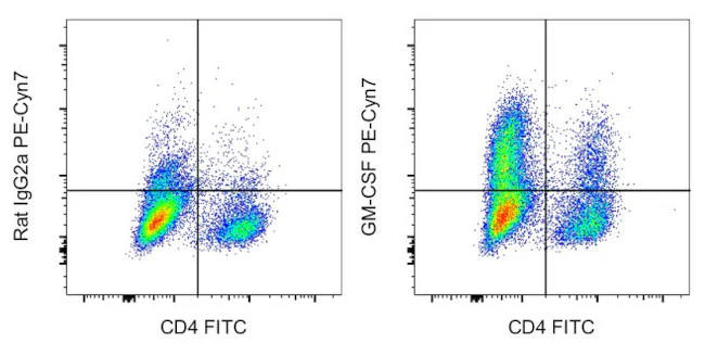
Applications Tested: This MP1-22E9 antibody has been tested by intracellular staining followed by flow cytometric analysis of restimulated mouse splenocytes. This may be used at less than or equal to 0.25 µg per test. A test is defined as the amount (µg) of antibody that will stain a cell sample in a final volume of 100 µL. Cell number should be determined empirically but can range from 10^5 to 10^8 cells/test. It is recommended that the antibody be carefully titrated for optimal performance in the assay of interest.
Light sensitivity: This tandem dye is sensitive to photo-induced oxidation. Please protect this vial and stained samples from light.
Fixation: Samples can be stored in IC Fixation Buffer (Product # 00-8222-49) (100 µL of cell sample + 100 µL of IC Fixation Buffer) or 1-step Fix/Lyse Solution (Product # 00-5333-57) for up to 3 days in the dark at 4°C with minimal impact on brightness and FRET efficiency/compensation. Some generalizations regarding fluorophore performance after fixation can be made, but clone specific performance should be determined empirically.
Excitation: 488-561 nm; Emission: 775 nm; Laser: Blue Laser, Green Laser, Yellow-Green Laser.
For Research Use Only. Not for use in diagnostic procedures. Not for resale without express authorization.
GM-CSF Monoclonal Antibody (MP1-22E9), PE-Cyanine7, eBioscience
PRODUCT DETAILS
Host: Rat
Isotype: IgG2a, kappa
Clonality: Monoclonal
Clone: MP1-22E9
Format: PE-Cyanine7
Reactivity: Ms
Application: Flow Cytometry
Tested Dilution: 0.25 µg/test
Concentration: 0.2 mg/mL
Storage: 4°C, store in dark, DO NOT FREEZE!
Formulation: PBS with 0.09% sodium azide; pH 7.2
Purification: Affinity chromatography
Data Sheet: TDS
Specific Information
Description: The MP1-22E9 antibody reacts with mouse granulocyte/macrophage - colony stimulating factor (GM-CSF). The MP1-22E9 antibody is a neutralizing antibody. mouse GM-CSF is a 14 kDa factor produced mainly by activated T cells and macrophages. Other cell types, such as endothelium and fibroblasts, also secrete GM-CSF in response to TNF-alpha, IL-2, IL-1, and IFN-gamma. GM-CSF stimulates growth of macrophages, granulocytes and dendritic cells. GM-CSF is found as a membrane-bound form and also as a complex associated with the extracellular matrix. Non-glycosylated GM-CSF is biologically active.
Applications Reported: This MP1-22E9 antibody has been reported for use in intracellular staining followed by flow cytometric analysis.
Applications Tested: This MP1-22E9 antibody has been tested by intracellular staining followed by flow cytometric analysis of restimulated mouse splenocytes. This may be used at less than or equal to 0.25 µg per test. A test is defined as the amount (µg) of antibody that will stain a cell sample in a final volume of 100 µL. Cell number should be determined empirically but can range from 10^5 to 10^8 cells/test. It is recommended that the antibody be carefully titrated for optimal performance in the assay of interest.
Light sensitivity: This tandem dye is sensitive to photo-induced oxidation. Please protect this vial and stained samples from light.
Fixation: Samples can be stored in IC Fixation Buffer (Product # 00-8222-49) (100 µL of cell sample + 100 µL of IC Fixation Buffer) or 1-step Fix/Lyse Solution (Product # 00-5333-57) for up to 3 days in the dark at 4°C with minimal impact on brightness and FRET efficiency/compensation. Some generalizations regarding fluorophore performance after fixation can be made, but clone specific performance should be determined empirically.
Excitation: 488-561 nm; Emission: 775 nm; Laser: Blue Laser, Green Laser, Yellow-Green Laser.
For Research Use Only. Not for use in diagnostic procedures. Not for resale without express authorization.
Product Information
Product Information
Shipping & Returns
Shipping & Returns
Description
PRODUCT DETAILS
Host: Rat
Isotype: IgG2a, kappa
Clonality: Monoclonal
Clone: MP1-22E9
Format: PE-Cyanine7
Reactivity: Ms
Application: Flow Cytometry
Tested Dilution: 0.25 µg/test
Concentration: 0.2 mg/mL
Storage: 4°C, store in dark, DO NOT FREEZE!
Formulation: PBS with 0.09% sodium azide; pH 7.2
Purification: Affinity chromatography
Data Sheet: TDS
Specific Information
Description: The MP1-22E9 antibody reacts with mouse granulocyte/macrophage - colony stimulating factor (GM-CSF). The MP1-22E9 antibody is a neutralizing antibody. mouse GM-CSF is a 14 kDa factor produced mainly by activated T cells and macrophages. Other cell types, such as endothelium and fibroblasts, also secrete GM-CSF in response to TNF-alpha, IL-2, IL-1, and IFN-gamma. GM-CSF stimulates growth of macrophages, granulocytes and dendritic cells. GM-CSF is found as a membrane-bound form and also as a complex associated with the extracellular matrix. Non-glycosylated GM-CSF is biologically active.
Applications Reported: This MP1-22E9 antibody has been reported for use in intracellular staining followed by flow cytometric analysis.
Applications Tested: This MP1-22E9 antibody has been tested by intracellular staining followed by flow cytometric analysis of restimulated mouse splenocytes. This may be used at less than or equal to 0.25 µg per test. A test is defined as the amount (µg) of antibody that will stain a cell sample in a final volume of 100 µL. Cell number should be determined empirically but can range from 10^5 to 10^8 cells/test. It is recommended that the antibody be carefully titrated for optimal performance in the assay of interest.
Light sensitivity: This tandem dye is sensitive to photo-induced oxidation. Please protect this vial and stained samples from light.
Fixation: Samples can be stored in IC Fixation Buffer (Product # 00-8222-49) (100 µL of cell sample + 100 µL of IC Fixation Buffer) or 1-step Fix/Lyse Solution (Product # 00-5333-57) for up to 3 days in the dark at 4°C with minimal impact on brightness and FRET efficiency/compensation. Some generalizations regarding fluorophore performance after fixation can be made, but clone specific performance should be determined empirically.
Excitation: 488-561 nm; Emission: 775 nm; Laser: Blue Laser, Green Laser, Yellow-Green Laser.
For Research Use Only. Not for use in diagnostic procedures. Not for resale without express authorization.


