Gata-3 Monoclonal Antibody (TWAJ), PE, eBioscience
PRODUCT DETAILS
Host: Rat
Isotype: IgG2b, kappa
Clonality: Monoclonal
Clone: TWAJ
Format: PE
Reactivity: Hu, Ms, Po, Rh
Application: Flow Cytometry
Tested Dilution: 5 µL (0.06 µg)/test
Concentration: 5 μL/Test
Storage: 4°C, store in dark, DO NOT FREEZE!
Formulation: PBS with BSA and 0.09% sodium azide; pH 7.2
Purification: Affinity chromatography
Data Sheet: TDS
Specific Information
Description: The monoclonal antibody TWAJ recognizes mouse and human Gata-3, a member of the Gata family of transcription factors. Gata-3 is a T cell-specific transcription factor important for thymic development and Th2 differentiation. Expression during embryonic development is found in the central nervous system, skin, mammary glands and kidney. During development, the expression of Gata-3 is essential as homozygous knock-out of Gata-3 is embryonic lethal. The Gata-3 is also essential for T cell commitment and survival. In the thymus, expression is found mainly on the CD4 single positive cells. During Th2 differentiation, Gata-3 binds to the IL-4 promoter as well as represses the expression of T-bet, thus inhibiting Th1 differentiation.
Alternative splice variants have been reported especially in the MCF7 cell line. The TWAJ Human/Mouse Gata-3 antibody will recognize both forms (50 and 45 kDa) of the protein.
Staining with the TWAJ Human/Mouse Gata-3 antibody requires the use of the Foxp3/Transcription Factor Staining Buffer Set.(Product # 00-5523-00)
Cross reactivity in rhesus monkeys has been published.
Applications Reported: This TWAJ antibody has been reported for use in intracellular staining followed by flow cytometric analysis.
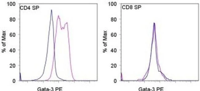
Applications Tested: This TWAJ antibody has been pre-titrated and tested by intracellular staining and flow cytometric analysis of mouse thymocytes using the Foxp3/Transcription Factor Staining Buffer Set (Product # 00-5523) and protocol. Please see BestProtocols® Section (Staining Intracellular Antigens for Flow Cytometry) for staining protocol (refer to Protocol B: One-step protocol for intracellular (nuclear) proteins). This can be used at 5 µL (0.06 µg) per test. A test is defined as the amount (µg) of antibody that will stain a cell sample in a final volume of 100 µL. Cell number should be determined empirically but can range from 10^5 to 10^8 cells/test.
Excitation: 488-561 nm; Emission: 578 nm; Laser: Blue Laser, Green Laser, Yellow-Green Laser.
Filtration: 0.2 µm post-manufacturing filtered.
For Research Use Only. Not for use in diagnostic procedures. Not for resale without express authorization.
Gata-3 Monoclonal Antibody (TWAJ), PE, eBioscience
PRODUCT DETAILS
Host: Rat
Isotype: IgG2b, kappa
Clonality: Monoclonal
Clone: TWAJ
Format: PE
Reactivity: Hu, Ms, Po, Rh
Application: Flow Cytometry
Tested Dilution: 5 µL (0.06 µg)/test
Concentration: 5 μL/Test
Storage: 4°C, store in dark, DO NOT FREEZE!
Formulation: PBS with BSA and 0.09% sodium azide; pH 7.2
Purification: Affinity chromatography
Data Sheet: TDS
Specific Information
Description: The monoclonal antibody TWAJ recognizes mouse and human Gata-3, a member of the Gata family of transcription factors. Gata-3 is a T cell-specific transcription factor important for thymic development and Th2 differentiation. Expression during embryonic development is found in the central nervous system, skin, mammary glands and kidney. During development, the expression of Gata-3 is essential as homozygous knock-out of Gata-3 is embryonic lethal. The Gata-3 is also essential for T cell commitment and survival. In the thymus, expression is found mainly on the CD4 single positive cells. During Th2 differentiation, Gata-3 binds to the IL-4 promoter as well as represses the expression of T-bet, thus inhibiting Th1 differentiation.
Alternative splice variants have been reported especially in the MCF7 cell line. The TWAJ Human/Mouse Gata-3 antibody will recognize both forms (50 and 45 kDa) of the protein.
Staining with the TWAJ Human/Mouse Gata-3 antibody requires the use of the Foxp3/Transcription Factor Staining Buffer Set.(Product # 00-5523-00)
Cross reactivity in rhesus monkeys has been published.
Applications Reported: This TWAJ antibody has been reported for use in intracellular staining followed by flow cytometric analysis.
Applications Tested: This TWAJ antibody has been pre-titrated and tested by intracellular staining and flow cytometric analysis of mouse thymocytes using the Foxp3/Transcription Factor Staining Buffer Set (Product # 00-5523) and protocol. Please see BestProtocols® Section (Staining Intracellular Antigens for Flow Cytometry) for staining protocol (refer to Protocol B: One-step protocol for intracellular (nuclear) proteins). This can be used at 5 µL (0.06 µg) per test. A test is defined as the amount (µg) of antibody that will stain a cell sample in a final volume of 100 µL. Cell number should be determined empirically but can range from 10^5 to 10^8 cells/test.
Excitation: 488-561 nm; Emission: 578 nm; Laser: Blue Laser, Green Laser, Yellow-Green Laser.
Filtration: 0.2 µm post-manufacturing filtered.
For Research Use Only. Not for use in diagnostic procedures. Not for resale without express authorization.
Product Information
Product Information
Shipping & Returns
Shipping & Returns
Description
PRODUCT DETAILS
Host: Rat
Isotype: IgG2b, kappa
Clonality: Monoclonal
Clone: TWAJ
Format: PE
Reactivity: Hu, Ms, Po, Rh
Application: Flow Cytometry
Tested Dilution: 5 µL (0.06 µg)/test
Concentration: 5 μL/Test
Storage: 4°C, store in dark, DO NOT FREEZE!
Formulation: PBS with BSA and 0.09% sodium azide; pH 7.2
Purification: Affinity chromatography
Data Sheet: TDS
Specific Information
Description: The monoclonal antibody TWAJ recognizes mouse and human Gata-3, a member of the Gata family of transcription factors. Gata-3 is a T cell-specific transcription factor important for thymic development and Th2 differentiation. Expression during embryonic development is found in the central nervous system, skin, mammary glands and kidney. During development, the expression of Gata-3 is essential as homozygous knock-out of Gata-3 is embryonic lethal. The Gata-3 is also essential for T cell commitment and survival. In the thymus, expression is found mainly on the CD4 single positive cells. During Th2 differentiation, Gata-3 binds to the IL-4 promoter as well as represses the expression of T-bet, thus inhibiting Th1 differentiation.
Alternative splice variants have been reported especially in the MCF7 cell line. The TWAJ Human/Mouse Gata-3 antibody will recognize both forms (50 and 45 kDa) of the protein.
Staining with the TWAJ Human/Mouse Gata-3 antibody requires the use of the Foxp3/Transcription Factor Staining Buffer Set.(Product # 00-5523-00)
Cross reactivity in rhesus monkeys has been published.
Applications Reported: This TWAJ antibody has been reported for use in intracellular staining followed by flow cytometric analysis.
Applications Tested: This TWAJ antibody has been pre-titrated and tested by intracellular staining and flow cytometric analysis of mouse thymocytes using the Foxp3/Transcription Factor Staining Buffer Set (Product # 00-5523) and protocol. Please see BestProtocols® Section (Staining Intracellular Antigens for Flow Cytometry) for staining protocol (refer to Protocol B: One-step protocol for intracellular (nuclear) proteins). This can be used at 5 µL (0.06 µg) per test. A test is defined as the amount (µg) of antibody that will stain a cell sample in a final volume of 100 µL. Cell number should be determined empirically but can range from 10^5 to 10^8 cells/test.
Excitation: 488-561 nm; Emission: 578 nm; Laser: Blue Laser, Green Laser, Yellow-Green Laser.
Filtration: 0.2 µm post-manufacturing filtered.
For Research Use Only. Not for use in diagnostic procedures. Not for resale without express authorization.


