FR4 Monoclonal Antibody (eBio12A5), PE, eBioscience
PRODUCT DETAILS
Host: Rat
Isotype: IgG2b, kappa
Clonality: Monoclonal
Clone: eBio12A5
Format: PE
Reactivity: Ms
Application: Flow Cytometry
Tested Dilution: 0.5 µg/test
Concentration: 0.2 mg/mL
Storage: 4°C, store in dark, DO NOT FREEZE!
Formulation: PBS with 0.09% sodium azide; pH 7.2
Purification: Affinity chromatography
Data Sheet: TDS
Specific Information
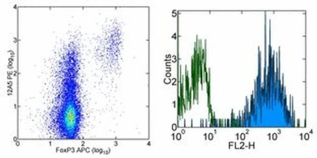
Description: The monoclonal antibody eBio12A5 recognizes FR4, also known as Folate receptor 4, FR delta and FBP3 (folate binding protein3). FR4 is a heavily glycosylated, 35 kD receptor for folic acid and the physiologic circulating form of the vitamin, N5-methyltetrahydrofolate. Natural Treg have high levels of FR4 and together with CD25 can be used to distinguish natural Treg, effector T cells, memory-like T cells and naive T cells. FR4 high CD25 high cells are natural Treg with high levels of Foxp3. FR4 high CD25 low cells are identified as central memory T cells, which upon stimulation, can proliferate and produce large amounts of IL-2. In contrast, FR4 low CD25 high cells secrete proinflammatory cytokines such as IFN gamma and IL-17 and have been characterized as effector memory T cells.
Based on co-staining studies, the epitopes recognized by eBioTH6 and eBio12A5 are different, thereby allowing functional studies with eBioTH6 to be evaluated with eBio12A5.
Applications Reported: This eBio12A5 antibody has been reported for use in flow cytometric analysis.
Applications Tested: This eBio12A5 antibody has been tested by flow cytometric analysis of mouse splenocytes. This can be used at less than or equal to 0.5 µg per test. A test is defined as the amount (µg) of antibody that will stain a cell sample in a final volume of 100 µL. Cell number should be determined empirically but can range from 10^5 to 10^8 cells/test. It is recommended that the antibody be carefully titrated for optimal performance in the assay of interest.
Excitation: 488-561 nm; Emission: 578 nm; Laser: Blue Laser, Green Laser, Yellow-Green Laser.
Filtration: 0.2 µm post-manufacturing filtered.
For Research Use Only. Not for use in diagnostic procedures. Not for resale without express authorization.
FR4 Monoclonal Antibody (eBio12A5), PE, eBioscience
PRODUCT DETAILS
Host: Rat
Isotype: IgG2b, kappa
Clonality: Monoclonal
Clone: eBio12A5
Format: PE
Reactivity: Ms
Application: Flow Cytometry
Tested Dilution: 0.5 µg/test
Concentration: 0.2 mg/mL
Storage: 4°C, store in dark, DO NOT FREEZE!
Formulation: PBS with 0.09% sodium azide; pH 7.2
Purification: Affinity chromatography
Data Sheet: TDS
Specific Information
Description: The monoclonal antibody eBio12A5 recognizes FR4, also known as Folate receptor 4, FR delta and FBP3 (folate binding protein3). FR4 is a heavily glycosylated, 35 kD receptor for folic acid and the physiologic circulating form of the vitamin, N5-methyltetrahydrofolate. Natural Treg have high levels of FR4 and together with CD25 can be used to distinguish natural Treg, effector T cells, memory-like T cells and naive T cells. FR4 high CD25 high cells are natural Treg with high levels of Foxp3. FR4 high CD25 low cells are identified as central memory T cells, which upon stimulation, can proliferate and produce large amounts of IL-2. In contrast, FR4 low CD25 high cells secrete proinflammatory cytokines such as IFN gamma and IL-17 and have been characterized as effector memory T cells.
Based on co-staining studies, the epitopes recognized by eBioTH6 and eBio12A5 are different, thereby allowing functional studies with eBioTH6 to be evaluated with eBio12A5.
Applications Reported: This eBio12A5 antibody has been reported for use in flow cytometric analysis.
Applications Tested: This eBio12A5 antibody has been tested by flow cytometric analysis of mouse splenocytes. This can be used at less than or equal to 0.5 µg per test. A test is defined as the amount (µg) of antibody that will stain a cell sample in a final volume of 100 µL. Cell number should be determined empirically but can range from 10^5 to 10^8 cells/test. It is recommended that the antibody be carefully titrated for optimal performance in the assay of interest.
Excitation: 488-561 nm; Emission: 578 nm; Laser: Blue Laser, Green Laser, Yellow-Green Laser.
Filtration: 0.2 µm post-manufacturing filtered.
For Research Use Only. Not for use in diagnostic procedures. Not for resale without express authorization.
Product Information
Product Information
Shipping & Returns
Shipping & Returns
Description
PRODUCT DETAILS
Host: Rat
Isotype: IgG2b, kappa
Clonality: Monoclonal
Clone: eBio12A5
Format: PE
Reactivity: Ms
Application: Flow Cytometry
Tested Dilution: 0.5 µg/test
Concentration: 0.2 mg/mL
Storage: 4°C, store in dark, DO NOT FREEZE!
Formulation: PBS with 0.09% sodium azide; pH 7.2
Purification: Affinity chromatography
Data Sheet: TDS
Specific Information
Description: The monoclonal antibody eBio12A5 recognizes FR4, also known as Folate receptor 4, FR delta and FBP3 (folate binding protein3). FR4 is a heavily glycosylated, 35 kD receptor for folic acid and the physiologic circulating form of the vitamin, N5-methyltetrahydrofolate. Natural Treg have high levels of FR4 and together with CD25 can be used to distinguish natural Treg, effector T cells, memory-like T cells and naive T cells. FR4 high CD25 high cells are natural Treg with high levels of Foxp3. FR4 high CD25 low cells are identified as central memory T cells, which upon stimulation, can proliferate and produce large amounts of IL-2. In contrast, FR4 low CD25 high cells secrete proinflammatory cytokines such as IFN gamma and IL-17 and have been characterized as effector memory T cells.
Based on co-staining studies, the epitopes recognized by eBioTH6 and eBio12A5 are different, thereby allowing functional studies with eBioTH6 to be evaluated with eBio12A5.
Applications Reported: This eBio12A5 antibody has been reported for use in flow cytometric analysis.
Applications Tested: This eBio12A5 antibody has been tested by flow cytometric analysis of mouse splenocytes. This can be used at less than or equal to 0.5 µg per test. A test is defined as the amount (µg) of antibody that will stain a cell sample in a final volume of 100 µL. Cell number should be determined empirically but can range from 10^5 to 10^8 cells/test. It is recommended that the antibody be carefully titrated for optimal performance in the assay of interest.
Excitation: 488-561 nm; Emission: 578 nm; Laser: Blue Laser, Green Laser, Yellow-Green Laser.
Filtration: 0.2 µm post-manufacturing filtered.
For Research Use Only. Not for use in diagnostic procedures. Not for resale without express authorization.


