FOXP3 Monoclonal Antibody (PCH101), PE-eFluor 610, eBioscience
PRODUCT DETAILS
Host: Rat
Isotype: IgG2a, kappa
Clonality: Monoclonal
Clone: PCH101
Format: PE-eFluor 610
Reactivity: Cp, Cy, Hu, Nhp, Rh
Application: Flow Cytometry
Tested Dilution: 5 µL (0.25 µg)/test
Concentration: 5 μL/Test
Storage: 4°C, store in dark, DO NOT FREEZE!
Formulation: PBS with BSA and 0.09% sodium azide; pH 7.2
Purification: Affinity chromatography
Data Sheet: TDS
Specific Information
Description: eBioscience offers a panel of monoclonal antibodies to different epitopes of human Foxp3, providing useful tools for investigating the complete expression pattern of Foxp3 at the protein level, and discerning the precise subsets of Foxp3^+ cells.
The PCH101 antibody reacts with the amino terminus of human foxp3 protein also known as FORKHEAD BOX P3, SCURFIN, and JM2; cross reactivity of this antibody to other proteins has not been determined. Foxp3, a 49-55 kDa protein, is a member of the forkhead/winged-helix family of transcriptional regulators, and was identified as the gene defective in 'scurfy' (sf) mice. Constitutive high expression of Foxp3 mRNA has been shown in CD4+CD25+ regulatory T cells (Treg cells), and ectopic expression of foxp3 in CD4+CD25- cells imparts a Treg phenotype in these cells.
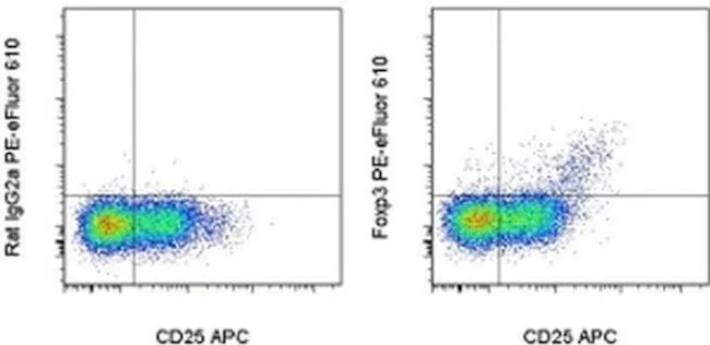
Intracellular staining of human peripheral blood mononuclear cells (PBMCs) with PCH101 antibody using the anti-human Foxp3 Staining Set and protocol reveals approximately 0.5-4% of lymphocytes staining, with the majority of staining occurring in the CD25^bright population. This is subject to donor variability.
PCH101 cross-reacts with rhesus, chimpanzee and cynomolgus. We recommend the use of CD4 (OKT4, Product # 11-0048-42 , or RPA-T4, Product # 11-0049-42 , depending on the species) and CD25 (BC96, Product # 17-0259-42).
Applications Reported: This PCH101 antibody has been reported for use in intracellular staining followed by flow cytometric analysis.
Applications Tested: This PCH101 antibody has been pre-titrated and tested by intracellular staining and flow cytometric analysis of normal human peripheral blood cells using the Foxp3/Transcription Factor Staining Buffer Set () and protocol. Refer to BestProtocols® for Staining Protocol (refer to Protocol B: One-step protocol for intracellular (nuclear) proteins). This can be used at 5 µL (0.25 µg) per test. A test is defined as the amount (µg) of antibody that will stain a cell sample in a final volume of 100 µL. Cell number should be determined empirically but can range from 10^5 to 10^8 cells/test.
PE-eFluor® 610 can be excited with laser lines from 488-561 nm and emits at 607 nm. We recommend using a 610/20 band pass filter (equivalent to PE-Texas Red®). Please make sure that your instrument is capable of detecting this fluorochrome.
Light sensitivity: This tandem dye is sensitive to photo-induced oxidation. Please protect this vial and stained samples from light.
Fixation: Samples can be stored in IC Fixation Buffer (Product # 00-8222) (100 µL of cell sample + 100 µL of IC Fixation Buffer) or 1-step Fix/Lyse Solution (Product # 00-5333) for up to 3 days in the dark at 4°C with minimal impact on brightness and FRET efficiency/compensation. Some generalizations regarding fluorophore performance after fixation can be made, but clone specific performance should be determined empirically.
Excitation: 488-561 nm; Emission: 607 nm; Laser: Blue Laser, Green Laser, Yellow-Green Laser.
Filtration: 0.2 µm post-manufacturing filtered.
For Research Use Only. Not for use in diagnostic procedures. Not for resale without express authorization.
FOXP3 Monoclonal Antibody (PCH101), PE-eFluor 610, eBioscience
PRODUCT DETAILS
Host: Rat
Isotype: IgG2a, kappa
Clonality: Monoclonal
Clone: PCH101
Format: PE-eFluor 610
Reactivity: Cp, Cy, Hu, Nhp, Rh
Application: Flow Cytometry
Tested Dilution: 5 µL (0.25 µg)/test
Concentration: 5 μL/Test
Storage: 4°C, store in dark, DO NOT FREEZE!
Formulation: PBS with BSA and 0.09% sodium azide; pH 7.2
Purification: Affinity chromatography
Data Sheet: TDS
Specific Information
Description: eBioscience offers a panel of monoclonal antibodies to different epitopes of human Foxp3, providing useful tools for investigating the complete expression pattern of Foxp3 at the protein level, and discerning the precise subsets of Foxp3^+ cells.
The PCH101 antibody reacts with the amino terminus of human foxp3 protein also known as FORKHEAD BOX P3, SCURFIN, and JM2; cross reactivity of this antibody to other proteins has not been determined. Foxp3, a 49-55 kDa protein, is a member of the forkhead/winged-helix family of transcriptional regulators, and was identified as the gene defective in 'scurfy' (sf) mice. Constitutive high expression of Foxp3 mRNA has been shown in CD4+CD25+ regulatory T cells (Treg cells), and ectopic expression of foxp3 in CD4+CD25- cells imparts a Treg phenotype in these cells.
Intracellular staining of human peripheral blood mononuclear cells (PBMCs) with PCH101 antibody using the anti-human Foxp3 Staining Set and protocol reveals approximately 0.5-4% of lymphocytes staining, with the majority of staining occurring in the CD25^bright population. This is subject to donor variability.
PCH101 cross-reacts with rhesus, chimpanzee and cynomolgus. We recommend the use of CD4 (OKT4, Product # 11-0048-42 , or RPA-T4, Product # 11-0049-42 , depending on the species) and CD25 (BC96, Product # 17-0259-42).
Applications Reported: This PCH101 antibody has been reported for use in intracellular staining followed by flow cytometric analysis.
Applications Tested: This PCH101 antibody has been pre-titrated and tested by intracellular staining and flow cytometric analysis of normal human peripheral blood cells using the Foxp3/Transcription Factor Staining Buffer Set () and protocol. Refer to BestProtocols® for Staining Protocol (refer to Protocol B: One-step protocol for intracellular (nuclear) proteins). This can be used at 5 µL (0.25 µg) per test. A test is defined as the amount (µg) of antibody that will stain a cell sample in a final volume of 100 µL. Cell number should be determined empirically but can range from 10^5 to 10^8 cells/test.
PE-eFluor® 610 can be excited with laser lines from 488-561 nm and emits at 607 nm. We recommend using a 610/20 band pass filter (equivalent to PE-Texas Red®). Please make sure that your instrument is capable of detecting this fluorochrome.
Light sensitivity: This tandem dye is sensitive to photo-induced oxidation. Please protect this vial and stained samples from light.
Fixation: Samples can be stored in IC Fixation Buffer (Product # 00-8222) (100 µL of cell sample + 100 µL of IC Fixation Buffer) or 1-step Fix/Lyse Solution (Product # 00-5333) for up to 3 days in the dark at 4°C with minimal impact on brightness and FRET efficiency/compensation. Some generalizations regarding fluorophore performance after fixation can be made, but clone specific performance should be determined empirically.
Excitation: 488-561 nm; Emission: 607 nm; Laser: Blue Laser, Green Laser, Yellow-Green Laser.
Filtration: 0.2 µm post-manufacturing filtered.
For Research Use Only. Not for use in diagnostic procedures. Not for resale without express authorization.
Product Information
Product Information
Shipping & Returns
Shipping & Returns
Description
PRODUCT DETAILS
Host: Rat
Isotype: IgG2a, kappa
Clonality: Monoclonal
Clone: PCH101
Format: PE-eFluor 610
Reactivity: Cp, Cy, Hu, Nhp, Rh
Application: Flow Cytometry
Tested Dilution: 5 µL (0.25 µg)/test
Concentration: 5 μL/Test
Storage: 4°C, store in dark, DO NOT FREEZE!
Formulation: PBS with BSA and 0.09% sodium azide; pH 7.2
Purification: Affinity chromatography
Data Sheet: TDS
Specific Information
Description: eBioscience offers a panel of monoclonal antibodies to different epitopes of human Foxp3, providing useful tools for investigating the complete expression pattern of Foxp3 at the protein level, and discerning the precise subsets of Foxp3^+ cells.
The PCH101 antibody reacts with the amino terminus of human foxp3 protein also known as FORKHEAD BOX P3, SCURFIN, and JM2; cross reactivity of this antibody to other proteins has not been determined. Foxp3, a 49-55 kDa protein, is a member of the forkhead/winged-helix family of transcriptional regulators, and was identified as the gene defective in 'scurfy' (sf) mice. Constitutive high expression of Foxp3 mRNA has been shown in CD4+CD25+ regulatory T cells (Treg cells), and ectopic expression of foxp3 in CD4+CD25- cells imparts a Treg phenotype in these cells.
Intracellular staining of human peripheral blood mononuclear cells (PBMCs) with PCH101 antibody using the anti-human Foxp3 Staining Set and protocol reveals approximately 0.5-4% of lymphocytes staining, with the majority of staining occurring in the CD25^bright population. This is subject to donor variability.
PCH101 cross-reacts with rhesus, chimpanzee and cynomolgus. We recommend the use of CD4 (OKT4, Product # 11-0048-42 , or RPA-T4, Product # 11-0049-42 , depending on the species) and CD25 (BC96, Product # 17-0259-42).
Applications Reported: This PCH101 antibody has been reported for use in intracellular staining followed by flow cytometric analysis.
Applications Tested: This PCH101 antibody has been pre-titrated and tested by intracellular staining and flow cytometric analysis of normal human peripheral blood cells using the Foxp3/Transcription Factor Staining Buffer Set () and protocol. Refer to BestProtocols® for Staining Protocol (refer to Protocol B: One-step protocol for intracellular (nuclear) proteins). This can be used at 5 µL (0.25 µg) per test. A test is defined as the amount (µg) of antibody that will stain a cell sample in a final volume of 100 µL. Cell number should be determined empirically but can range from 10^5 to 10^8 cells/test.
PE-eFluor® 610 can be excited with laser lines from 488-561 nm and emits at 607 nm. We recommend using a 610/20 band pass filter (equivalent to PE-Texas Red®). Please make sure that your instrument is capable of detecting this fluorochrome.
Light sensitivity: This tandem dye is sensitive to photo-induced oxidation. Please protect this vial and stained samples from light.
Fixation: Samples can be stored in IC Fixation Buffer (Product # 00-8222) (100 µL of cell sample + 100 µL of IC Fixation Buffer) or 1-step Fix/Lyse Solution (Product # 00-5333) for up to 3 days in the dark at 4°C with minimal impact on brightness and FRET efficiency/compensation. Some generalizations regarding fluorophore performance after fixation can be made, but clone specific performance should be determined empirically.
Excitation: 488-561 nm; Emission: 607 nm; Laser: Blue Laser, Green Laser, Yellow-Green Laser.
Filtration: 0.2 µm post-manufacturing filtered.
For Research Use Only. Not for use in diagnostic procedures. Not for resale without express authorization.


