FOXP3 Monoclonal Antibody (NRRF-30), PE, eBioscience
PRODUCT DETAILS
Host: Rat
Isotype: IgG2a, kappa
Clonality: Monoclonal
Clone: NRRF-30
Format: PE
Reactivity: Ms
Application: Flow Cytometry
Tested Dilution: 0.25 µg/test
Concentration: 0.2 mg/mL
Storage: 4°C, store in dark, DO NOT FREEZE!
Formulation: PBS with 0.09% sodium azide; pH 7.2
Purification: Affinity chromatography
Data Sheet: TDS
Specific Information
Description: The NRRF-30 antibody reacts with mouse Foxp3 also known as FORKHEAD BOX P3, SCURFIN, and JM2; cross reactivity of this antibody to other proteins has not been determined. Foxp3, a 49-55 kDa protein, is a member of the forkhead/winged-helix family of transcriptional regulators, and was identified as the gene defective in 'scurfy' (sf) mice. Constitutive high expression of foxP3 mRNA has been shown in CD4+/CD25+ regulatory T cells (Treg cells), and ectopic expression of foxp3 in CD4+/CD25- cells imparts a Treg phenotype in these cells.
Immunoblotting with NRRF-30 antibody has mapped the epitope to amino acids 1-75 of the mouse Foxp3 protein.
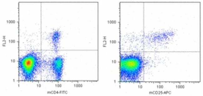
Intracellular staining of mouse splenocytes with fluorochrome-conjugated NRRF-30 using the eBioscience Foxp3 Staining Buffers () and corresponding staining protocol reveals approximately 3% of total cells in the C57Bl/6 strain and approximately 5% in the BALB/c mouse strain. Multicolor flow cytometric analysis demonstrates approximately 90% of the CD4+/CD2+ cells and 4% of the CD4+/CD25- cells staining with NRRF-30. Co-staining with FJK-16s (anti-mouse/rat Foxp3, Product # 71-5775-40), which has been mapped to amino acids 71-125, and NRRF-30 shows 100% correlation, indicating that the same cells are stained with both anti-mouse Foxp3 antibodies.
Please see our FAQ regarding the usage of eBioscience Foxp3 reagents.
Applications Reported: This NRRF-30 antibody has been reported for use in intracellular staining followed by flow cytometric analysis.
Applications Tested: This NRRF-30 antibody has been tested by intracellular flow cytometric analysis of mouse splenocytes using the Foxp3/Transcription Factor Staining Buffer Set (Product # 00-5523-00) and protocol. Please see BestProtocols® Section (Staining Intracellular Antigens for Flow Cytometry) for staining protocol (refer to Protocol B: One-step protocol for intracellular (nuclear) proteins). This can be used at less than or equal to 0.25 µg per test. A test is defined as the amount (µg) of antibody that will stain a cell sample in a final volume of 100 µL. Cell number should be determined empirically but can range from 10^5 to 10^8 cells/test. It is recommended that the antibody be carefully titrated for optimal performance in the assay of interest.
Excitation: 488-561 nm; Emission: 578 nm; Laser: Blue Laser, Green Laser, Yellow-Green Laser.
Filtration: 0.2 µm post-manufacturing filtered.
For Research Use Only. Not for use in diagnostic procedures. Not for resale without express authorization.
Original: $520.00
-70%$520.00
$156.00FOXP3 Monoclonal Antibody (NRRF-30), PE, eBioscience
PRODUCT DETAILS
Host: Rat
Isotype: IgG2a, kappa
Clonality: Monoclonal
Clone: NRRF-30
Format: PE
Reactivity: Ms
Application: Flow Cytometry
Tested Dilution: 0.25 µg/test
Concentration: 0.2 mg/mL
Storage: 4°C, store in dark, DO NOT FREEZE!
Formulation: PBS with 0.09% sodium azide; pH 7.2
Purification: Affinity chromatography
Data Sheet: TDS
Specific Information
Description: The NRRF-30 antibody reacts with mouse Foxp3 also known as FORKHEAD BOX P3, SCURFIN, and JM2; cross reactivity of this antibody to other proteins has not been determined. Foxp3, a 49-55 kDa protein, is a member of the forkhead/winged-helix family of transcriptional regulators, and was identified as the gene defective in 'scurfy' (sf) mice. Constitutive high expression of foxP3 mRNA has been shown in CD4+/CD25+ regulatory T cells (Treg cells), and ectopic expression of foxp3 in CD4+/CD25- cells imparts a Treg phenotype in these cells.
Immunoblotting with NRRF-30 antibody has mapped the epitope to amino acids 1-75 of the mouse Foxp3 protein.
Intracellular staining of mouse splenocytes with fluorochrome-conjugated NRRF-30 using the eBioscience Foxp3 Staining Buffers () and corresponding staining protocol reveals approximately 3% of total cells in the C57Bl/6 strain and approximately 5% in the BALB/c mouse strain. Multicolor flow cytometric analysis demonstrates approximately 90% of the CD4+/CD2+ cells and 4% of the CD4+/CD25- cells staining with NRRF-30. Co-staining with FJK-16s (anti-mouse/rat Foxp3, Product # 71-5775-40), which has been mapped to amino acids 71-125, and NRRF-30 shows 100% correlation, indicating that the same cells are stained with both anti-mouse Foxp3 antibodies.
Please see our FAQ regarding the usage of eBioscience Foxp3 reagents.
Applications Reported: This NRRF-30 antibody has been reported for use in intracellular staining followed by flow cytometric analysis.
Applications Tested: This NRRF-30 antibody has been tested by intracellular flow cytometric analysis of mouse splenocytes using the Foxp3/Transcription Factor Staining Buffer Set (Product # 00-5523-00) and protocol. Please see BestProtocols® Section (Staining Intracellular Antigens for Flow Cytometry) for staining protocol (refer to Protocol B: One-step protocol for intracellular (nuclear) proteins). This can be used at less than or equal to 0.25 µg per test. A test is defined as the amount (µg) of antibody that will stain a cell sample in a final volume of 100 µL. Cell number should be determined empirically but can range from 10^5 to 10^8 cells/test. It is recommended that the antibody be carefully titrated for optimal performance in the assay of interest.
Excitation: 488-561 nm; Emission: 578 nm; Laser: Blue Laser, Green Laser, Yellow-Green Laser.
Filtration: 0.2 µm post-manufacturing filtered.
For Research Use Only. Not for use in diagnostic procedures. Not for resale without express authorization.
Product Information
Product Information
Shipping & Returns
Shipping & Returns
Description
PRODUCT DETAILS
Host: Rat
Isotype: IgG2a, kappa
Clonality: Monoclonal
Clone: NRRF-30
Format: PE
Reactivity: Ms
Application: Flow Cytometry
Tested Dilution: 0.25 µg/test
Concentration: 0.2 mg/mL
Storage: 4°C, store in dark, DO NOT FREEZE!
Formulation: PBS with 0.09% sodium azide; pH 7.2
Purification: Affinity chromatography
Data Sheet: TDS
Specific Information
Description: The NRRF-30 antibody reacts with mouse Foxp3 also known as FORKHEAD BOX P3, SCURFIN, and JM2; cross reactivity of this antibody to other proteins has not been determined. Foxp3, a 49-55 kDa protein, is a member of the forkhead/winged-helix family of transcriptional regulators, and was identified as the gene defective in 'scurfy' (sf) mice. Constitutive high expression of foxP3 mRNA has been shown in CD4+/CD25+ regulatory T cells (Treg cells), and ectopic expression of foxp3 in CD4+/CD25- cells imparts a Treg phenotype in these cells.
Immunoblotting with NRRF-30 antibody has mapped the epitope to amino acids 1-75 of the mouse Foxp3 protein.
Intracellular staining of mouse splenocytes with fluorochrome-conjugated NRRF-30 using the eBioscience Foxp3 Staining Buffers () and corresponding staining protocol reveals approximately 3% of total cells in the C57Bl/6 strain and approximately 5% in the BALB/c mouse strain. Multicolor flow cytometric analysis demonstrates approximately 90% of the CD4+/CD2+ cells and 4% of the CD4+/CD25- cells staining with NRRF-30. Co-staining with FJK-16s (anti-mouse/rat Foxp3, Product # 71-5775-40), which has been mapped to amino acids 71-125, and NRRF-30 shows 100% correlation, indicating that the same cells are stained with both anti-mouse Foxp3 antibodies.
Please see our FAQ regarding the usage of eBioscience Foxp3 reagents.
Applications Reported: This NRRF-30 antibody has been reported for use in intracellular staining followed by flow cytometric analysis.
Applications Tested: This NRRF-30 antibody has been tested by intracellular flow cytometric analysis of mouse splenocytes using the Foxp3/Transcription Factor Staining Buffer Set (Product # 00-5523-00) and protocol. Please see BestProtocols® Section (Staining Intracellular Antigens for Flow Cytometry) for staining protocol (refer to Protocol B: One-step protocol for intracellular (nuclear) proteins). This can be used at less than or equal to 0.25 µg per test. A test is defined as the amount (µg) of antibody that will stain a cell sample in a final volume of 100 µL. Cell number should be determined empirically but can range from 10^5 to 10^8 cells/test. It is recommended that the antibody be carefully titrated for optimal performance in the assay of interest.
Excitation: 488-561 nm; Emission: 578 nm; Laser: Blue Laser, Green Laser, Yellow-Green Laser.
Filtration: 0.2 µm post-manufacturing filtered.
For Research Use Only. Not for use in diagnostic procedures. Not for resale without express authorization.


