F4/80 Monoclonal Antibody (BM8), Alexa Fluor 561, eBioscience
PRODUCT DETAILS
Host: Rat
Isotype: IgG2a, kappa
Clonality: Monoclonal
Clone: BM8
Format: Alexa Fluor 561
Reactivity: Ms
Application: Flow Cytometry
Tested Dilution: 0.25 µg/test
Concentration: 0.2 mg/mL
Storage: 4°C, store in dark, DO NOT FREEZE!
Formulation: PBS with 0.09% sodium azide; pH 7.2
Purification: Affinity chromatography
Data Sheet: TDS
Specific Information
Description: The BM8 monoclonal antibody reacts with mouse F4/80 antigen, an approximately 160 kDa surface receptor. It belongs to the EGF-TM7 family of proteins. As such it contains seven EGF-like domains on its extracellular N-terminus, seven transmembrane spanning sequences, and an intracellular C-terminal domain showing homology to other TM7 superfamily members. The F4/80 antigen is expressed by a majority of mature macrophages and is one of the best markers for this population of cells. However, other cell types, such as peritoneal eosinophils, Langerhans cells, and some other dendritic cell subtypes, have been reported to express this antigen as well. Expression of F4/80 commences during early myeloid development in vivo and can be upregulated on BM cells stimulated in vitro with M-CSF. Some populations of macrophages, especially in the lymphoid microenvironment, may be devoid of F4/80.
Applications Reported: This BM8 antibody has been reported for use in flow cytometric analysis.
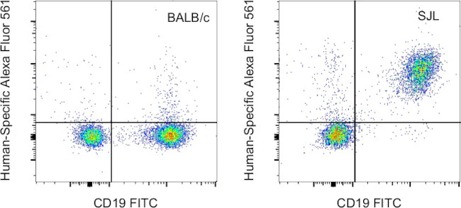
Our internal testing shows that Alexa Fluor 561 non-specifically stains B cells in Swiss Webster and SJL mice. Non-specific staining has not been observed in BALB/c or C57BL/6 mice. Other strains have not been tested. See the Antibody Testing Data for an example of this strain-dependent difference.
Applications Tested: This BM8 antibody has been tested by flow cytometric analysis of mouse resident peritoneal macrophages. This may be used at less than or equal to 0.25 µg per test. A test is defined as the amount (µg) of antibody that will stain a cell sample in a final volume of 100 µL. Cell number should be determined empirically but can range from 10^5 to 10^8 cells/test. It is recommended that the antibody be carefully titrated for optimal performance in the assay of interest.
Alexa Fluor 561 emits at 575 nm and is intended for use on spectral cytometers where it may be multiplexed with both PE and PE-eFluor 610.
Excitation: 558 nm; Emission: 575 nm; Laser: Yellow-Green Laser
For Research Use Only. Not for use in diagnostic procedures. Not for resale without express authorization.
F4/80 Monoclonal Antibody (BM8), Alexa Fluor 561, eBioscience
PRODUCT DETAILS
Host: Rat
Isotype: IgG2a, kappa
Clonality: Monoclonal
Clone: BM8
Format: Alexa Fluor 561
Reactivity: Ms
Application: Flow Cytometry
Tested Dilution: 0.25 µg/test
Concentration: 0.2 mg/mL
Storage: 4°C, store in dark, DO NOT FREEZE!
Formulation: PBS with 0.09% sodium azide; pH 7.2
Purification: Affinity chromatography
Data Sheet: TDS
Specific Information
Description: The BM8 monoclonal antibody reacts with mouse F4/80 antigen, an approximately 160 kDa surface receptor. It belongs to the EGF-TM7 family of proteins. As such it contains seven EGF-like domains on its extracellular N-terminus, seven transmembrane spanning sequences, and an intracellular C-terminal domain showing homology to other TM7 superfamily members. The F4/80 antigen is expressed by a majority of mature macrophages and is one of the best markers for this population of cells. However, other cell types, such as peritoneal eosinophils, Langerhans cells, and some other dendritic cell subtypes, have been reported to express this antigen as well. Expression of F4/80 commences during early myeloid development in vivo and can be upregulated on BM cells stimulated in vitro with M-CSF. Some populations of macrophages, especially in the lymphoid microenvironment, may be devoid of F4/80.
Applications Reported: This BM8 antibody has been reported for use in flow cytometric analysis.
Our internal testing shows that Alexa Fluor 561 non-specifically stains B cells in Swiss Webster and SJL mice. Non-specific staining has not been observed in BALB/c or C57BL/6 mice. Other strains have not been tested. See the Antibody Testing Data for an example of this strain-dependent difference.
Applications Tested: This BM8 antibody has been tested by flow cytometric analysis of mouse resident peritoneal macrophages. This may be used at less than or equal to 0.25 µg per test. A test is defined as the amount (µg) of antibody that will stain a cell sample in a final volume of 100 µL. Cell number should be determined empirically but can range from 10^5 to 10^8 cells/test. It is recommended that the antibody be carefully titrated for optimal performance in the assay of interest.
Alexa Fluor 561 emits at 575 nm and is intended for use on spectral cytometers where it may be multiplexed with both PE and PE-eFluor 610.
Excitation: 558 nm; Emission: 575 nm; Laser: Yellow-Green Laser
For Research Use Only. Not for use in diagnostic procedures. Not for resale without express authorization.
Product Information
Product Information
Shipping & Returns
Shipping & Returns
Description
PRODUCT DETAILS
Host: Rat
Isotype: IgG2a, kappa
Clonality: Monoclonal
Clone: BM8
Format: Alexa Fluor 561
Reactivity: Ms
Application: Flow Cytometry
Tested Dilution: 0.25 µg/test
Concentration: 0.2 mg/mL
Storage: 4°C, store in dark, DO NOT FREEZE!
Formulation: PBS with 0.09% sodium azide; pH 7.2
Purification: Affinity chromatography
Data Sheet: TDS
Specific Information
Description: The BM8 monoclonal antibody reacts with mouse F4/80 antigen, an approximately 160 kDa surface receptor. It belongs to the EGF-TM7 family of proteins. As such it contains seven EGF-like domains on its extracellular N-terminus, seven transmembrane spanning sequences, and an intracellular C-terminal domain showing homology to other TM7 superfamily members. The F4/80 antigen is expressed by a majority of mature macrophages and is one of the best markers for this population of cells. However, other cell types, such as peritoneal eosinophils, Langerhans cells, and some other dendritic cell subtypes, have been reported to express this antigen as well. Expression of F4/80 commences during early myeloid development in vivo and can be upregulated on BM cells stimulated in vitro with M-CSF. Some populations of macrophages, especially in the lymphoid microenvironment, may be devoid of F4/80.
Applications Reported: This BM8 antibody has been reported for use in flow cytometric analysis.
Our internal testing shows that Alexa Fluor 561 non-specifically stains B cells in Swiss Webster and SJL mice. Non-specific staining has not been observed in BALB/c or C57BL/6 mice. Other strains have not been tested. See the Antibody Testing Data for an example of this strain-dependent difference.
Applications Tested: This BM8 antibody has been tested by flow cytometric analysis of mouse resident peritoneal macrophages. This may be used at less than or equal to 0.25 µg per test. A test is defined as the amount (µg) of antibody that will stain a cell sample in a final volume of 100 µL. Cell number should be determined empirically but can range from 10^5 to 10^8 cells/test. It is recommended that the antibody be carefully titrated for optimal performance in the assay of interest.
Alexa Fluor 561 emits at 575 nm and is intended for use on spectral cytometers where it may be multiplexed with both PE and PE-eFluor 610.
Excitation: 558 nm; Emission: 575 nm; Laser: Yellow-Green Laser
For Research Use Only. Not for use in diagnostic procedures. Not for resale without express authorization.


