EGR1 Monoclonal Antibody (HEGR1DS), PE, eBioscience
PRODUCT DETAILS
Host: Rat
Isotype: IgG2a, kappa
Clonality: Monoclonal
Clone: HEGR1DS
Format: PE
Reactivity: Hu
Application: Flow Cytometry
Tested Dilution: 5 µL (0.03 µg)/test
Concentration: 5 μL/Test
Storage: 4°C, store in dark, DO NOT FREEZE!
Formulation: PBS with BSA and 0.09% sodium azide; pH 7.2
Purification: Affinity chromatography
Data Sheet: TDS
Specific Information
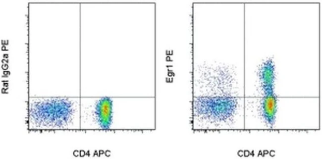
Description: The monoclonal antibody HEGR1DS recognizes human Egr1 (Early Growth Response 1), a C2H2-type zinc-finger transcription factor broadly expressed in various types of tissues. Egr1 is induced within minutes after cell activation and it regulates expression of numerous factors involved in cell division, growth, and differentiation. It has been shown to suppress tumorigenesis by inducing apoptosis of cancer cells but it can also promote their proliferation. Egr1 is often used as an early cell activation marker of T and B lymphocytes as well as myeloid cells.
Applications Reported: This HEGR1DS antibody has been reported for use in intracellular staining followed by flow cytometric analysis.
Applications Tested: This HEGR1DS antibody has been pre-titrated and tested by intracellular staining and flow cytometric analysis of stimulated normal human peripheral blood cells using the Foxp3/Transcription Factor Staining Buffer Set (Product # 00-5523-00) and protocol. Please refer to BestProtocols®: Protocol B: One step protocol for (nuclear) intracellular proteins located under the Resources Tab online. The use of the Intracellular Fixation & Permeabilization Buffer Set (Product # 88-8824-00) and protocol is not recommended. This antibody can be used at 5 µL (0.03 µg) per test. A test is defined as the amount (µg) of antibody that will stain a cell sample in a final volume of 100 µL. Cell number should be determined empirically but can range from 10^5 to 10^8 cells/test.
Excitation: 488-561 nm; Emission: 578 nm; Laser: Blue Laser, Green Laser, Yellow-Green Laser.
Filtration: 0.2 µm post-manufacturing filtered.
For Research Use Only. Not for use in diagnostic procedures. Not for resale without express authorization.
EGR1 Monoclonal Antibody (HEGR1DS), PE, eBioscience
PRODUCT DETAILS
Host: Rat
Isotype: IgG2a, kappa
Clonality: Monoclonal
Clone: HEGR1DS
Format: PE
Reactivity: Hu
Application: Flow Cytometry
Tested Dilution: 5 µL (0.03 µg)/test
Concentration: 5 μL/Test
Storage: 4°C, store in dark, DO NOT FREEZE!
Formulation: PBS with BSA and 0.09% sodium azide; pH 7.2
Purification: Affinity chromatography
Data Sheet: TDS
Specific Information
Description: The monoclonal antibody HEGR1DS recognizes human Egr1 (Early Growth Response 1), a C2H2-type zinc-finger transcription factor broadly expressed in various types of tissues. Egr1 is induced within minutes after cell activation and it regulates expression of numerous factors involved in cell division, growth, and differentiation. It has been shown to suppress tumorigenesis by inducing apoptosis of cancer cells but it can also promote their proliferation. Egr1 is often used as an early cell activation marker of T and B lymphocytes as well as myeloid cells.
Applications Reported: This HEGR1DS antibody has been reported for use in intracellular staining followed by flow cytometric analysis.
Applications Tested: This HEGR1DS antibody has been pre-titrated and tested by intracellular staining and flow cytometric analysis of stimulated normal human peripheral blood cells using the Foxp3/Transcription Factor Staining Buffer Set (Product # 00-5523-00) and protocol. Please refer to BestProtocols®: Protocol B: One step protocol for (nuclear) intracellular proteins located under the Resources Tab online. The use of the Intracellular Fixation & Permeabilization Buffer Set (Product # 88-8824-00) and protocol is not recommended. This antibody can be used at 5 µL (0.03 µg) per test. A test is defined as the amount (µg) of antibody that will stain a cell sample in a final volume of 100 µL. Cell number should be determined empirically but can range from 10^5 to 10^8 cells/test.
Excitation: 488-561 nm; Emission: 578 nm; Laser: Blue Laser, Green Laser, Yellow-Green Laser.
Filtration: 0.2 µm post-manufacturing filtered.
For Research Use Only. Not for use in diagnostic procedures. Not for resale without express authorization.
Product Information
Product Information
Shipping & Returns
Shipping & Returns
Description
PRODUCT DETAILS
Host: Rat
Isotype: IgG2a, kappa
Clonality: Monoclonal
Clone: HEGR1DS
Format: PE
Reactivity: Hu
Application: Flow Cytometry
Tested Dilution: 5 µL (0.03 µg)/test
Concentration: 5 μL/Test
Storage: 4°C, store in dark, DO NOT FREEZE!
Formulation: PBS with BSA and 0.09% sodium azide; pH 7.2
Purification: Affinity chromatography
Data Sheet: TDS
Specific Information
Description: The monoclonal antibody HEGR1DS recognizes human Egr1 (Early Growth Response 1), a C2H2-type zinc-finger transcription factor broadly expressed in various types of tissues. Egr1 is induced within minutes after cell activation and it regulates expression of numerous factors involved in cell division, growth, and differentiation. It has been shown to suppress tumorigenesis by inducing apoptosis of cancer cells but it can also promote their proliferation. Egr1 is often used as an early cell activation marker of T and B lymphocytes as well as myeloid cells.
Applications Reported: This HEGR1DS antibody has been reported for use in intracellular staining followed by flow cytometric analysis.
Applications Tested: This HEGR1DS antibody has been pre-titrated and tested by intracellular staining and flow cytometric analysis of stimulated normal human peripheral blood cells using the Foxp3/Transcription Factor Staining Buffer Set (Product # 00-5523-00) and protocol. Please refer to BestProtocols®: Protocol B: One step protocol for (nuclear) intracellular proteins located under the Resources Tab online. The use of the Intracellular Fixation & Permeabilization Buffer Set (Product # 88-8824-00) and protocol is not recommended. This antibody can be used at 5 µL (0.03 µg) per test. A test is defined as the amount (µg) of antibody that will stain a cell sample in a final volume of 100 µL. Cell number should be determined empirically but can range from 10^5 to 10^8 cells/test.
Excitation: 488-561 nm; Emission: 578 nm; Laser: Blue Laser, Green Laser, Yellow-Green Laser.
Filtration: 0.2 µm post-manufacturing filtered.
For Research Use Only. Not for use in diagnostic procedures. Not for resale without express authorization.


