EGFR Monoclonal Antibody (me1B3), eFluor 660, eBioscience
PRODUCT DETAILS
Host: Mouse
Isotype: IgG1, kappa
Clonality: Monoclonal
Clone: me1B3
Format: eFluor 660
Reactivity: Hu
Application: Flow Cytometry
Tested Dilution: 5 µL (0.125 µg)/test
Concentration: 5 μL/Test
Storage: 4°C, store in dark, DO NOT FREEZE!
Formulation: PBS with BSA and 0.09% sodium azide; pH 7.2
Purification: Affinity chromatography
Data Sheet: TDS
Specific Information
Description: This me1B3 monoclonal antibody reacts with human epidermal growth factor receptor (EGFR), also known as ErbB1 and Her1. EGFR is a receptor tyrosine kinase belonging to the EGFR/ErbB receptor subfamily, which also includes ErbB2/Her2/neu. EGFR contains an extracellular ligand-binding domain, a single transmembrane-spanning region, and a cytoplasmic tail containing a tyrosine kinase domain. Specific ligands include EGF and TNF alpha, as well as other members of the EGF protein family. Upon ligand binding, EGFR undergoes receptor-mediated dimerization with itself or ErbB2/Her2/neu. Dimerization induces auto-phosphorylation, resulting in the activation of PLC gamma, JAK/STAT, MAPK/ERK, and PI3K/AKT signaling pathways. EGFR has a role in cell proliferation, migration and differentiation, and is evolutionarily highly conserved in structure and function. EGFR is broadly expressed and is overexpressed or mutated in glioblastomas and a number of different epithelial cancers, including non-small cell lung cancer and colorectal cancer.
Applications Reported: This me1B3 antibody has been reported for use in flow cytometric analysis.
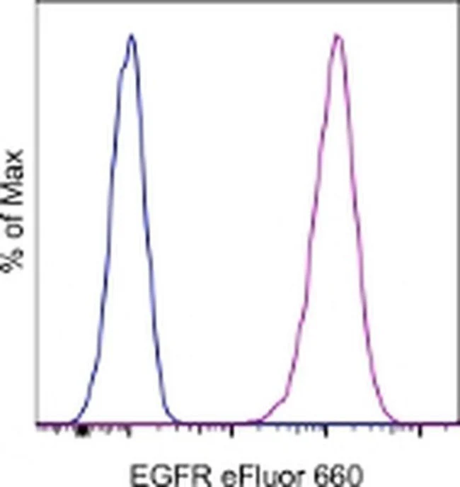
Applications Tested: This me1B3 antibody has been pre-titrated and tested by flow cytometric analysis of HeLa cells. This can be used at 5 µL (0.125 µg) per test. A test is defined as the amount (µg) of antibody that will stain a cell sample in a final volume of 100 µL. Cell number should be determined empirically but can range from 10^5 to 10^8 cells/test.
eFluor™ 660 is a replacement for Alexa Fluor™ 647. eFluor™ 660 emits at 659 nm and is excited with the red laser (633 nm). Please make sure that your instrument is capable of detecting this fluorochome.
Excitation: 633-647 nm; Emission: 668 nm; Laser: Red Laser.
Filtration: 0.2 µm post-manufacturing filtered.
For Research Use Only. Not for use in diagnostic procedures. Not for resale without express authorization.
EGFR Monoclonal Antibody (me1B3), eFluor 660, eBioscience
PRODUCT DETAILS
Host: Mouse
Isotype: IgG1, kappa
Clonality: Monoclonal
Clone: me1B3
Format: eFluor 660
Reactivity: Hu
Application: Flow Cytometry
Tested Dilution: 5 µL (0.125 µg)/test
Concentration: 5 μL/Test
Storage: 4°C, store in dark, DO NOT FREEZE!
Formulation: PBS with BSA and 0.09% sodium azide; pH 7.2
Purification: Affinity chromatography
Data Sheet: TDS
Specific Information
Description: This me1B3 monoclonal antibody reacts with human epidermal growth factor receptor (EGFR), also known as ErbB1 and Her1. EGFR is a receptor tyrosine kinase belonging to the EGFR/ErbB receptor subfamily, which also includes ErbB2/Her2/neu. EGFR contains an extracellular ligand-binding domain, a single transmembrane-spanning region, and a cytoplasmic tail containing a tyrosine kinase domain. Specific ligands include EGF and TNF alpha, as well as other members of the EGF protein family. Upon ligand binding, EGFR undergoes receptor-mediated dimerization with itself or ErbB2/Her2/neu. Dimerization induces auto-phosphorylation, resulting in the activation of PLC gamma, JAK/STAT, MAPK/ERK, and PI3K/AKT signaling pathways. EGFR has a role in cell proliferation, migration and differentiation, and is evolutionarily highly conserved in structure and function. EGFR is broadly expressed and is overexpressed or mutated in glioblastomas and a number of different epithelial cancers, including non-small cell lung cancer and colorectal cancer.
Applications Reported: This me1B3 antibody has been reported for use in flow cytometric analysis.
Applications Tested: This me1B3 antibody has been pre-titrated and tested by flow cytometric analysis of HeLa cells. This can be used at 5 µL (0.125 µg) per test. A test is defined as the amount (µg) of antibody that will stain a cell sample in a final volume of 100 µL. Cell number should be determined empirically but can range from 10^5 to 10^8 cells/test.
eFluor™ 660 is a replacement for Alexa Fluor™ 647. eFluor™ 660 emits at 659 nm and is excited with the red laser (633 nm). Please make sure that your instrument is capable of detecting this fluorochome.
Excitation: 633-647 nm; Emission: 668 nm; Laser: Red Laser.
Filtration: 0.2 µm post-manufacturing filtered.
For Research Use Only. Not for use in diagnostic procedures. Not for resale without express authorization.
Product Information
Product Information
Shipping & Returns
Shipping & Returns
Description
PRODUCT DETAILS
Host: Mouse
Isotype: IgG1, kappa
Clonality: Monoclonal
Clone: me1B3
Format: eFluor 660
Reactivity: Hu
Application: Flow Cytometry
Tested Dilution: 5 µL (0.125 µg)/test
Concentration: 5 μL/Test
Storage: 4°C, store in dark, DO NOT FREEZE!
Formulation: PBS with BSA and 0.09% sodium azide; pH 7.2
Purification: Affinity chromatography
Data Sheet: TDS
Specific Information
Description: This me1B3 monoclonal antibody reacts with human epidermal growth factor receptor (EGFR), also known as ErbB1 and Her1. EGFR is a receptor tyrosine kinase belonging to the EGFR/ErbB receptor subfamily, which also includes ErbB2/Her2/neu. EGFR contains an extracellular ligand-binding domain, a single transmembrane-spanning region, and a cytoplasmic tail containing a tyrosine kinase domain. Specific ligands include EGF and TNF alpha, as well as other members of the EGF protein family. Upon ligand binding, EGFR undergoes receptor-mediated dimerization with itself or ErbB2/Her2/neu. Dimerization induces auto-phosphorylation, resulting in the activation of PLC gamma, JAK/STAT, MAPK/ERK, and PI3K/AKT signaling pathways. EGFR has a role in cell proliferation, migration and differentiation, and is evolutionarily highly conserved in structure and function. EGFR is broadly expressed and is overexpressed or mutated in glioblastomas and a number of different epithelial cancers, including non-small cell lung cancer and colorectal cancer.
Applications Reported: This me1B3 antibody has been reported for use in flow cytometric analysis.
Applications Tested: This me1B3 antibody has been pre-titrated and tested by flow cytometric analysis of HeLa cells. This can be used at 5 µL (0.125 µg) per test. A test is defined as the amount (µg) of antibody that will stain a cell sample in a final volume of 100 µL. Cell number should be determined empirically but can range from 10^5 to 10^8 cells/test.
eFluor™ 660 is a replacement for Alexa Fluor™ 647. eFluor™ 660 emits at 659 nm and is excited with the red laser (633 nm). Please make sure that your instrument is capable of detecting this fluorochome.
Excitation: 633-647 nm; Emission: 668 nm; Laser: Red Laser.
Filtration: 0.2 µm post-manufacturing filtered.
For Research Use Only. Not for use in diagnostic procedures. Not for resale without express authorization.


