CX3CR1 Monoclonal Antibody (2A9-1), PE-Cyanine7, eBioscience
PRODUCT DETAILS
Host: Rat
Isotype: IgG2b, kappa
Clonality: Monoclonal
Clone: 2A9-1
Format: PE-Cyanine7
Reactivity: Hu
Application: Flow Cytometry
Tested Dilution: 5 µL (0.125 µg)/test
Concentration: 5 μL/Test
Storage: 4°C, store in dark, DO NOT FREEZE!
Formulation: PBS with BSA and 0.09% sodium azide; pH 7.2
Purification: Affinity chromatography
Data Sheet: TDS
Specific Information
Description: This 2A9-1 monoclonal antibody reacts with human CX3CR1, which is the receptor for fractalkine, a transmembrane chemokine of the CX3C family. CX3CR1 is a seven transmembrane G protein-coupled receptor expressed on CD16+ NK cells, T cells (e.g. CD8+, CD4+, and gamma/delta), and monocytes. In non-immune cells, CX3CR1 has been found on osteoclast precursors and microglia. Little to no CX3CR1 surface expression can be detected on B cells and granulocytes. Together, fractalkine and its receptor mediate cell-cell adhesion and chemotaxis of NK cells, T cells, and monocytes. The expression of CX3CR1 has also been correlated with high levels of intracellular perforin and granzyme B.
Applications Reported: This 2A9-1 antibody has been reported for use in flow cytometric analysis.
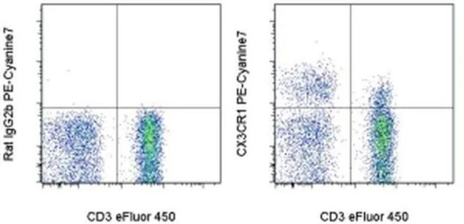
Applications Tested: This 2A9-1 antibody has been pre-titrated and tested by flow cytometric analysis of normal human peripheral blood cells. This can be used at 5 µL (0.125 µg) per test. A test is defined as the amount (µg) of antibody that will stain a cell sample in a final volume of 100 µL. Cell number should be determined empirically but can range from 10^5 to 10^8 cells/test.
Light sensitivity: This tandem dye is sensitive to photo-induced oxidation. Please protect this vial and stained samples from light.
Fixation: Samples can be stored in IC Fixation Buffer (Product # 00-822-49) (100 µL of cell sample + 100 µL of IC Fixation Buffer) or 1-step Fix/Lyse Solution (Product # 00-5333-54) for up to 3 days in the dark at 4°C with minimal impact on brightness and FRET efficiency/compensation. Some generalizations regarding fluorophore performance after fixation can be made, but clone specific performance should be determined empirically.
Excitation: 488-561 nm; Emission: 775 nm; Laser: Blue Laser, Green Laser, Yellow-Green Laser.
Filtration: 0.2 µm post-manufacturing filtered.
For Research Use Only. Not for use in diagnostic procedures. Not for resale without express authorization.
Original: $514.00
-70%$514.00
$154.20CX3CR1 Monoclonal Antibody (2A9-1), PE-Cyanine7, eBioscience
PRODUCT DETAILS
Host: Rat
Isotype: IgG2b, kappa
Clonality: Monoclonal
Clone: 2A9-1
Format: PE-Cyanine7
Reactivity: Hu
Application: Flow Cytometry
Tested Dilution: 5 µL (0.125 µg)/test
Concentration: 5 μL/Test
Storage: 4°C, store in dark, DO NOT FREEZE!
Formulation: PBS with BSA and 0.09% sodium azide; pH 7.2
Purification: Affinity chromatography
Data Sheet: TDS
Specific Information
Description: This 2A9-1 monoclonal antibody reacts with human CX3CR1, which is the receptor for fractalkine, a transmembrane chemokine of the CX3C family. CX3CR1 is a seven transmembrane G protein-coupled receptor expressed on CD16+ NK cells, T cells (e.g. CD8+, CD4+, and gamma/delta), and monocytes. In non-immune cells, CX3CR1 has been found on osteoclast precursors and microglia. Little to no CX3CR1 surface expression can be detected on B cells and granulocytes. Together, fractalkine and its receptor mediate cell-cell adhesion and chemotaxis of NK cells, T cells, and monocytes. The expression of CX3CR1 has also been correlated with high levels of intracellular perforin and granzyme B.
Applications Reported: This 2A9-1 antibody has been reported for use in flow cytometric analysis.
Applications Tested: This 2A9-1 antibody has been pre-titrated and tested by flow cytometric analysis of normal human peripheral blood cells. This can be used at 5 µL (0.125 µg) per test. A test is defined as the amount (µg) of antibody that will stain a cell sample in a final volume of 100 µL. Cell number should be determined empirically but can range from 10^5 to 10^8 cells/test.
Light sensitivity: This tandem dye is sensitive to photo-induced oxidation. Please protect this vial and stained samples from light.
Fixation: Samples can be stored in IC Fixation Buffer (Product # 00-822-49) (100 µL of cell sample + 100 µL of IC Fixation Buffer) or 1-step Fix/Lyse Solution (Product # 00-5333-54) for up to 3 days in the dark at 4°C with minimal impact on brightness and FRET efficiency/compensation. Some generalizations regarding fluorophore performance after fixation can be made, but clone specific performance should be determined empirically.
Excitation: 488-561 nm; Emission: 775 nm; Laser: Blue Laser, Green Laser, Yellow-Green Laser.
Filtration: 0.2 µm post-manufacturing filtered.
For Research Use Only. Not for use in diagnostic procedures. Not for resale without express authorization.
Product Information
Product Information
Shipping & Returns
Shipping & Returns
Description
PRODUCT DETAILS
Host: Rat
Isotype: IgG2b, kappa
Clonality: Monoclonal
Clone: 2A9-1
Format: PE-Cyanine7
Reactivity: Hu
Application: Flow Cytometry
Tested Dilution: 5 µL (0.125 µg)/test
Concentration: 5 μL/Test
Storage: 4°C, store in dark, DO NOT FREEZE!
Formulation: PBS with BSA and 0.09% sodium azide; pH 7.2
Purification: Affinity chromatography
Data Sheet: TDS
Specific Information
Description: This 2A9-1 monoclonal antibody reacts with human CX3CR1, which is the receptor for fractalkine, a transmembrane chemokine of the CX3C family. CX3CR1 is a seven transmembrane G protein-coupled receptor expressed on CD16+ NK cells, T cells (e.g. CD8+, CD4+, and gamma/delta), and monocytes. In non-immune cells, CX3CR1 has been found on osteoclast precursors and microglia. Little to no CX3CR1 surface expression can be detected on B cells and granulocytes. Together, fractalkine and its receptor mediate cell-cell adhesion and chemotaxis of NK cells, T cells, and monocytes. The expression of CX3CR1 has also been correlated with high levels of intracellular perforin and granzyme B.
Applications Reported: This 2A9-1 antibody has been reported for use in flow cytometric analysis.
Applications Tested: This 2A9-1 antibody has been pre-titrated and tested by flow cytometric analysis of normal human peripheral blood cells. This can be used at 5 µL (0.125 µg) per test. A test is defined as the amount (µg) of antibody that will stain a cell sample in a final volume of 100 µL. Cell number should be determined empirically but can range from 10^5 to 10^8 cells/test.
Light sensitivity: This tandem dye is sensitive to photo-induced oxidation. Please protect this vial and stained samples from light.
Fixation: Samples can be stored in IC Fixation Buffer (Product # 00-822-49) (100 µL of cell sample + 100 µL of IC Fixation Buffer) or 1-step Fix/Lyse Solution (Product # 00-5333-54) for up to 3 days in the dark at 4°C with minimal impact on brightness and FRET efficiency/compensation. Some generalizations regarding fluorophore performance after fixation can be made, but clone specific performance should be determined empirically.
Excitation: 488-561 nm; Emission: 775 nm; Laser: Blue Laser, Green Laser, Yellow-Green Laser.
Filtration: 0.2 µm post-manufacturing filtered.
For Research Use Only. Not for use in diagnostic procedures. Not for resale without express authorization.


