CD8b Monoclonal Antibody (eBioH35-17.2 (H35-17.2)), Super Bright 436, eBioscience
PRODUCT DETAILS
Host: Rat
Isotype: IgG2b, kappa
Clonality: Monoclonal
Clone: eBioH35-17.2 (H35-17.2)
Format: Super Bright 436
Reactivity: Ms
Application: Flow Cytometry
Tested Dilution: 0.25 µg/test
Concentration: 0.2 mg/mL
Storage: 4°C, store in dark, DO NOT FREEZE!
Formulation: PBS with BSA and 0.09% sodium azide; pH 7.2
Purification: Affinity chromatography
Data Sheet: TDS
Specific Information
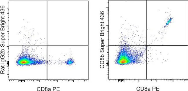
Description: The eBioH35-17.2 monoclonal antibody reacts with the mouse CD8 beta molecule. The CD8 beta chain associates with the CD8 alpha chain to form the CD8 alpha/beta heterodimer expressed on the surface of a majority of thymocytes, and on peripheral cytotoxic alpha beta TCR T cells. CD8 binds to MHC class I and plays a role in T cell development and activation of mature T cells.
Applications Reported: This eBioH35-17.2 (H35-17.2) antibody has been reported for use in flow cytometric analysis.
Applications Tested: This eBioH35-17.2 (H35-17.2) antibody has been tested by flow cytometric analysis of mouse splenocytes. This can be used at less than or equal to 0.25 µg per test. A test is defined as the amount (µg) of antibody that will stain a cell sample in a final volume of 100 µL. Cell number should be determined empirically but can range from 10^5 to 10^8 cells/test. It is recommended that the antibody be carefully titrated for optimal performance in the assay of interest.
Super Bright 436 can be excited with the violet laser line (405 nm) and emits at 436 nm. We recommend using a 450/50 bandpass filter, or equivalent. Please make sure that your instrument is capable of detecting this fluorochrome.
When using two or more Super Bright dye-conjugated antibodies in a staining panel, it is recommended to use Super Bright Complete Staining Buffer (Product # SB-4401) to minimize any non-specific polymer interactions. Please refer to the datasheet for Super Bright Staining Buffer for more information.
Excitation: 405 nm; Emission: 436 nm; Laser: Violet Laser
Super Bright Polymer Dyes are sold under license from Becton, Dickinson and Company.
For Research Use Only. Not for use in diagnostic procedures. Not for resale without express authorization.
Original: $392.00
-70%$392.00
$117.60CD8b Monoclonal Antibody (eBioH35-17.2 (H35-17.2)), Super Bright 436, eBioscience
PRODUCT DETAILS
Host: Rat
Isotype: IgG2b, kappa
Clonality: Monoclonal
Clone: eBioH35-17.2 (H35-17.2)
Format: Super Bright 436
Reactivity: Ms
Application: Flow Cytometry
Tested Dilution: 0.25 µg/test
Concentration: 0.2 mg/mL
Storage: 4°C, store in dark, DO NOT FREEZE!
Formulation: PBS with BSA and 0.09% sodium azide; pH 7.2
Purification: Affinity chromatography
Data Sheet: TDS
Specific Information
Description: The eBioH35-17.2 monoclonal antibody reacts with the mouse CD8 beta molecule. The CD8 beta chain associates with the CD8 alpha chain to form the CD8 alpha/beta heterodimer expressed on the surface of a majority of thymocytes, and on peripheral cytotoxic alpha beta TCR T cells. CD8 binds to MHC class I and plays a role in T cell development and activation of mature T cells.
Applications Reported: This eBioH35-17.2 (H35-17.2) antibody has been reported for use in flow cytometric analysis.
Applications Tested: This eBioH35-17.2 (H35-17.2) antibody has been tested by flow cytometric analysis of mouse splenocytes. This can be used at less than or equal to 0.25 µg per test. A test is defined as the amount (µg) of antibody that will stain a cell sample in a final volume of 100 µL. Cell number should be determined empirically but can range from 10^5 to 10^8 cells/test. It is recommended that the antibody be carefully titrated for optimal performance in the assay of interest.
Super Bright 436 can be excited with the violet laser line (405 nm) and emits at 436 nm. We recommend using a 450/50 bandpass filter, or equivalent. Please make sure that your instrument is capable of detecting this fluorochrome.
When using two or more Super Bright dye-conjugated antibodies in a staining panel, it is recommended to use Super Bright Complete Staining Buffer (Product # SB-4401) to minimize any non-specific polymer interactions. Please refer to the datasheet for Super Bright Staining Buffer for more information.
Excitation: 405 nm; Emission: 436 nm; Laser: Violet Laser
Super Bright Polymer Dyes are sold under license from Becton, Dickinson and Company.
For Research Use Only. Not for use in diagnostic procedures. Not for resale without express authorization.
Product Information
Product Information
Shipping & Returns
Shipping & Returns
Description
PRODUCT DETAILS
Host: Rat
Isotype: IgG2b, kappa
Clonality: Monoclonal
Clone: eBioH35-17.2 (H35-17.2)
Format: Super Bright 436
Reactivity: Ms
Application: Flow Cytometry
Tested Dilution: 0.25 µg/test
Concentration: 0.2 mg/mL
Storage: 4°C, store in dark, DO NOT FREEZE!
Formulation: PBS with BSA and 0.09% sodium azide; pH 7.2
Purification: Affinity chromatography
Data Sheet: TDS
Specific Information
Description: The eBioH35-17.2 monoclonal antibody reacts with the mouse CD8 beta molecule. The CD8 beta chain associates with the CD8 alpha chain to form the CD8 alpha/beta heterodimer expressed on the surface of a majority of thymocytes, and on peripheral cytotoxic alpha beta TCR T cells. CD8 binds to MHC class I and plays a role in T cell development and activation of mature T cells.
Applications Reported: This eBioH35-17.2 (H35-17.2) antibody has been reported for use in flow cytometric analysis.
Applications Tested: This eBioH35-17.2 (H35-17.2) antibody has been tested by flow cytometric analysis of mouse splenocytes. This can be used at less than or equal to 0.25 µg per test. A test is defined as the amount (µg) of antibody that will stain a cell sample in a final volume of 100 µL. Cell number should be determined empirically but can range from 10^5 to 10^8 cells/test. It is recommended that the antibody be carefully titrated for optimal performance in the assay of interest.
Super Bright 436 can be excited with the violet laser line (405 nm) and emits at 436 nm. We recommend using a 450/50 bandpass filter, or equivalent. Please make sure that your instrument is capable of detecting this fluorochrome.
When using two or more Super Bright dye-conjugated antibodies in a staining panel, it is recommended to use Super Bright Complete Staining Buffer (Product # SB-4401) to minimize any non-specific polymer interactions. Please refer to the datasheet for Super Bright Staining Buffer for more information.
Excitation: 405 nm; Emission: 436 nm; Laser: Violet Laser
Super Bright Polymer Dyes are sold under license from Becton, Dickinson and Company.
For Research Use Only. Not for use in diagnostic procedures. Not for resale without express authorization.


