CD8a Monoclonal Antibody (53-6.7), PE-Cyanine5.5, eBioscience
PRODUCT DETAILS
Host: Rat
Isotype: IgG2a, kappa
Clonality: Monoclonal
Clone: 53-6.7
Format: PE-Cyanine5.5
Reactivity: Ms
Application: Flow Cytometry
Tested Dilution: 0.125 µg/test
Concentration: 0.2 mg/mL
Storage: 4°C, store in dark, DO NOT FREEZE!
Formulation: PBS with 0.09% sodium azide; pH 7.2
Purification: Affinity chromatography
Data Sheet: TDS
Specific Information
Description: The 53-6.7 monoclonal antibody reacts with the mouse CD8a molecule. CD8a is an approximately 32-34 kDa cell surface receptor expressed either as a heterodimer with the CD8 beta chain (CD8 alpha beta) or as a homodimer (CD8 alpha alpha). A majority of thymocytes and a subpopulation of mature alpha beta TCR T cells express CD8 alpha beta while gamma delta TCR T cells, a subpopulation of intestinal intraepithelial lymphocytes (IELs) and dendritic cells express CD8 alpha alpha. CD8 binds to MHC class I and through its association with protein tyrosine kinase p56lck plays a role in T cell development and activation of mature T cells.
Applications Reported: This 53-6.7 antibody has been reported for use in flow cytometric analysis.
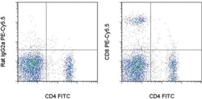
Applications Tested: This 53-6.7 antibody has been tested by flow cytometric analysis of mouse splenocytes. This can be used at less than or equal to 0.125 µg per test. A test is defined as the amount (µg) of antibody that will stain a cell sample in a final volume of 100 µL. Cell number should be determined empirically but can range from 10^5 to 10^8 cells/test. It is recommended that the antibody be carefully titrated for optimal performance in the assay of interest.
Light sensitivity: This tandem dye is sensitive photo-induced oxidation. Please protect this vial and stained samples from light.
Fixation: Samples can be stored in IC Fixation Buffer (Product # 00-822-49) (100 µL cell sample + 100 µL IC Fixation Buffer) or 1-step Fix/Lyse Solution (Product # 00-5333-54) for up to 3 days in the dark at 4°C with minimal impact on brightness and FRET efficiency/compensation. Some generalizations regarding fluorophore performance after fixation can be made, but clone specific performance should be determined empirically.
Excitation: 488-561 nm; Emission: 695 nm; Laser: Blue Laser, Green Laser, Yellow-Green Laser.
For Research Use Only. Not for use in diagnostic procedures. Not for resale without express authorization.
Original: $421.00
-70%$421.00
$126.30CD8a Monoclonal Antibody (53-6.7), PE-Cyanine5.5, eBioscience
PRODUCT DETAILS
Host: Rat
Isotype: IgG2a, kappa
Clonality: Monoclonal
Clone: 53-6.7
Format: PE-Cyanine5.5
Reactivity: Ms
Application: Flow Cytometry
Tested Dilution: 0.125 µg/test
Concentration: 0.2 mg/mL
Storage: 4°C, store in dark, DO NOT FREEZE!
Formulation: PBS with 0.09% sodium azide; pH 7.2
Purification: Affinity chromatography
Data Sheet: TDS
Specific Information
Description: The 53-6.7 monoclonal antibody reacts with the mouse CD8a molecule. CD8a is an approximately 32-34 kDa cell surface receptor expressed either as a heterodimer with the CD8 beta chain (CD8 alpha beta) or as a homodimer (CD8 alpha alpha). A majority of thymocytes and a subpopulation of mature alpha beta TCR T cells express CD8 alpha beta while gamma delta TCR T cells, a subpopulation of intestinal intraepithelial lymphocytes (IELs) and dendritic cells express CD8 alpha alpha. CD8 binds to MHC class I and through its association with protein tyrosine kinase p56lck plays a role in T cell development and activation of mature T cells.
Applications Reported: This 53-6.7 antibody has been reported for use in flow cytometric analysis.
Applications Tested: This 53-6.7 antibody has been tested by flow cytometric analysis of mouse splenocytes. This can be used at less than or equal to 0.125 µg per test. A test is defined as the amount (µg) of antibody that will stain a cell sample in a final volume of 100 µL. Cell number should be determined empirically but can range from 10^5 to 10^8 cells/test. It is recommended that the antibody be carefully titrated for optimal performance in the assay of interest.
Light sensitivity: This tandem dye is sensitive photo-induced oxidation. Please protect this vial and stained samples from light.
Fixation: Samples can be stored in IC Fixation Buffer (Product # 00-822-49) (100 µL cell sample + 100 µL IC Fixation Buffer) or 1-step Fix/Lyse Solution (Product # 00-5333-54) for up to 3 days in the dark at 4°C with minimal impact on brightness and FRET efficiency/compensation. Some generalizations regarding fluorophore performance after fixation can be made, but clone specific performance should be determined empirically.
Excitation: 488-561 nm; Emission: 695 nm; Laser: Blue Laser, Green Laser, Yellow-Green Laser.
For Research Use Only. Not for use in diagnostic procedures. Not for resale without express authorization.
Product Information
Product Information
Shipping & Returns
Shipping & Returns
Description
PRODUCT DETAILS
Host: Rat
Isotype: IgG2a, kappa
Clonality: Monoclonal
Clone: 53-6.7
Format: PE-Cyanine5.5
Reactivity: Ms
Application: Flow Cytometry
Tested Dilution: 0.125 µg/test
Concentration: 0.2 mg/mL
Storage: 4°C, store in dark, DO NOT FREEZE!
Formulation: PBS with 0.09% sodium azide; pH 7.2
Purification: Affinity chromatography
Data Sheet: TDS
Specific Information
Description: The 53-6.7 monoclonal antibody reacts with the mouse CD8a molecule. CD8a is an approximately 32-34 kDa cell surface receptor expressed either as a heterodimer with the CD8 beta chain (CD8 alpha beta) or as a homodimer (CD8 alpha alpha). A majority of thymocytes and a subpopulation of mature alpha beta TCR T cells express CD8 alpha beta while gamma delta TCR T cells, a subpopulation of intestinal intraepithelial lymphocytes (IELs) and dendritic cells express CD8 alpha alpha. CD8 binds to MHC class I and through its association with protein tyrosine kinase p56lck plays a role in T cell development and activation of mature T cells.
Applications Reported: This 53-6.7 antibody has been reported for use in flow cytometric analysis.
Applications Tested: This 53-6.7 antibody has been tested by flow cytometric analysis of mouse splenocytes. This can be used at less than or equal to 0.125 µg per test. A test is defined as the amount (µg) of antibody that will stain a cell sample in a final volume of 100 µL. Cell number should be determined empirically but can range from 10^5 to 10^8 cells/test. It is recommended that the antibody be carefully titrated for optimal performance in the assay of interest.
Light sensitivity: This tandem dye is sensitive photo-induced oxidation. Please protect this vial and stained samples from light.
Fixation: Samples can be stored in IC Fixation Buffer (Product # 00-822-49) (100 µL cell sample + 100 µL IC Fixation Buffer) or 1-step Fix/Lyse Solution (Product # 00-5333-54) for up to 3 days in the dark at 4°C with minimal impact on brightness and FRET efficiency/compensation. Some generalizations regarding fluorophore performance after fixation can be made, but clone specific performance should be determined empirically.
Excitation: 488-561 nm; Emission: 695 nm; Laser: Blue Laser, Green Laser, Yellow-Green Laser.
For Research Use Only. Not for use in diagnostic procedures. Not for resale without express authorization.


