CD85k (Gp49b) Monoclonal Antibody (H1.1), PE, eBioscience
PRODUCT DETAILS
Host: Armenian Hamster
Isotype: IgG
Clonality: Monoclonal
Clone: H1.1
Format: PE
Reactivity: Ms
Application: Flow Cytometry
Tested Dilution: 0.5 µg/test
Concentration: 0.2 mg/mL
Storage: 4°C, store in dark, DO NOT FREEZE!
Formulation: PBS with 0.09% sodium azide; pH 7.2
Purification: Affinity chromatography
Data Sheet: TDS
Specific Information
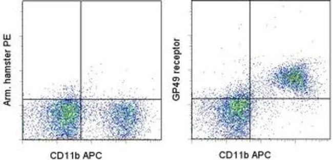
Description: The monoclonal antibody H1.1 recognizes gp49, a receptor with structural similarities to the PIR (paired Ig-activating receptors) and ILT/KIR(killer inhibitory receptors) family of proteins. Two closely linked genes on mouse chromosome 10 encode the related proteins gp49A and gp49B. These two receptors are almost 90% identical in their extracellular regions but differ in their cytoplasmic domains; gp49B contains an ITAM domain while gp49A does not and is extremely short. Expression of gp49 was originally identified on mast and LAK (IL-2 activated NK) cells. Expression on LAK cells has been shown to be predominantly gp49B with only a small amount of gp49A. Additionally gp49 is found on myeloid cells. Detailed function is not known but gp49B can inhibit cellular activation. The monoclonal antibody H1.1 recognizes both gp49A and gp49B.
Applications Reported: This H1.1 antibody has been reported for use in flow cytometric analysis.
Applications Tested: This H1.1 antibody has been tested by flow cytometric analysis of mouse bone marrow cells. This can be used at less than or equal to 0.5 µg per test. A test is defined as the amount (µg) of antibody that will stain a cell sample in a final volume of 100 µL. Cell number should be determined empirically but can range from 10^5 to 10^8 cells/test. It is recommended that the antibody be carefully titrated for optimal performance in the assay of interest.
Excitation: 488-561 nm; Emission: 578 nm; Laser: Blue Laser, Green Laser, Yellow-Green Laser.
Filtration: 0.2 µm post-manufacturing filtered.
For Research Use Only. Not for use in diagnostic procedures. Not for resale without express authorization.
CD85k (Gp49b) Monoclonal Antibody (H1.1), PE, eBioscience
PRODUCT DETAILS
Host: Armenian Hamster
Isotype: IgG
Clonality: Monoclonal
Clone: H1.1
Format: PE
Reactivity: Ms
Application: Flow Cytometry
Tested Dilution: 0.5 µg/test
Concentration: 0.2 mg/mL
Storage: 4°C, store in dark, DO NOT FREEZE!
Formulation: PBS with 0.09% sodium azide; pH 7.2
Purification: Affinity chromatography
Data Sheet: TDS
Specific Information
Description: The monoclonal antibody H1.1 recognizes gp49, a receptor with structural similarities to the PIR (paired Ig-activating receptors) and ILT/KIR(killer inhibitory receptors) family of proteins. Two closely linked genes on mouse chromosome 10 encode the related proteins gp49A and gp49B. These two receptors are almost 90% identical in their extracellular regions but differ in their cytoplasmic domains; gp49B contains an ITAM domain while gp49A does not and is extremely short. Expression of gp49 was originally identified on mast and LAK (IL-2 activated NK) cells. Expression on LAK cells has been shown to be predominantly gp49B with only a small amount of gp49A. Additionally gp49 is found on myeloid cells. Detailed function is not known but gp49B can inhibit cellular activation. The monoclonal antibody H1.1 recognizes both gp49A and gp49B.
Applications Reported: This H1.1 antibody has been reported for use in flow cytometric analysis.
Applications Tested: This H1.1 antibody has been tested by flow cytometric analysis of mouse bone marrow cells. This can be used at less than or equal to 0.5 µg per test. A test is defined as the amount (µg) of antibody that will stain a cell sample in a final volume of 100 µL. Cell number should be determined empirically but can range from 10^5 to 10^8 cells/test. It is recommended that the antibody be carefully titrated for optimal performance in the assay of interest.
Excitation: 488-561 nm; Emission: 578 nm; Laser: Blue Laser, Green Laser, Yellow-Green Laser.
Filtration: 0.2 µm post-manufacturing filtered.
For Research Use Only. Not for use in diagnostic procedures. Not for resale without express authorization.
Product Information
Product Information
Shipping & Returns
Shipping & Returns
Description
PRODUCT DETAILS
Host: Armenian Hamster
Isotype: IgG
Clonality: Monoclonal
Clone: H1.1
Format: PE
Reactivity: Ms
Application: Flow Cytometry
Tested Dilution: 0.5 µg/test
Concentration: 0.2 mg/mL
Storage: 4°C, store in dark, DO NOT FREEZE!
Formulation: PBS with 0.09% sodium azide; pH 7.2
Purification: Affinity chromatography
Data Sheet: TDS
Specific Information
Description: The monoclonal antibody H1.1 recognizes gp49, a receptor with structural similarities to the PIR (paired Ig-activating receptors) and ILT/KIR(killer inhibitory receptors) family of proteins. Two closely linked genes on mouse chromosome 10 encode the related proteins gp49A and gp49B. These two receptors are almost 90% identical in their extracellular regions but differ in their cytoplasmic domains; gp49B contains an ITAM domain while gp49A does not and is extremely short. Expression of gp49 was originally identified on mast and LAK (IL-2 activated NK) cells. Expression on LAK cells has been shown to be predominantly gp49B with only a small amount of gp49A. Additionally gp49 is found on myeloid cells. Detailed function is not known but gp49B can inhibit cellular activation. The monoclonal antibody H1.1 recognizes both gp49A and gp49B.
Applications Reported: This H1.1 antibody has been reported for use in flow cytometric analysis.
Applications Tested: This H1.1 antibody has been tested by flow cytometric analysis of mouse bone marrow cells. This can be used at less than or equal to 0.5 µg per test. A test is defined as the amount (µg) of antibody that will stain a cell sample in a final volume of 100 µL. Cell number should be determined empirically but can range from 10^5 to 10^8 cells/test. It is recommended that the antibody be carefully titrated for optimal performance in the assay of interest.
Excitation: 488-561 nm; Emission: 578 nm; Laser: Blue Laser, Green Laser, Yellow-Green Laser.
Filtration: 0.2 µm post-manufacturing filtered.
For Research Use Only. Not for use in diagnostic procedures. Not for resale without express authorization.


