CD79a Monoclonal Antibody (HM47), APC-eFluor 780, eBioscience
PRODUCT DETAILS
Host: Mouse
Isotype: IgG1, kappa
Clonality: Monoclonal
Clone: HM47
Format: APC-eFluor 780
Reactivity: Bb, Bv, Ca, Ck, Cp, Eq, GP, Hu, Ms, Nhp, Po, Rh, Rt
Application: Flow Cytometry
Tested Dilution: 5 µL (0.06 µg)/test
Concentration: 5 μL/Test
Storage: 4°C, store in dark, DO NOT FREEZE!
Formulation: PBS with BSA and 0.09% sodium azide; pH 7.2
Purification: Affinity chromatography
Data Sheet: TDS
Specific Information
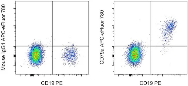
Description: The monoclonal antibody HM47 recognizes the cytoplasmic domain of CD79a, also known as mb-1. CD79a is a 47 kDa membrane glycoprotein that associates with CD79b to form the heterodimeric B cell receptor (BCR). This receptor is responsible for B cell signaling, resulting in activation, apoptosis or anergy. Expression of CD79a is found throughout development from the earliest pre-B cell to plasma cells. CD79 is expressed on B cell neoplasms and some non-B cell malignancies such as AML. Unlike other antibodies that can be masked in B cell neoplastic cells, HM47 is a reliable antibody for recognizing CD79a and, as such, is a good marker for B cells. Becauseseveral variants of CD79a exist, the exact location of the epitope may differ slightly. Nevertheless, in humans thie epitope is typically amino acids 208-222.
The HM47 antibody reacts to human, mouse, rat, dog, primates (rhesus, chimpanzee, macaque and baboon) pig, guinea rabbit, pig, horse, cow and chicken.
Applications Reported: This HM47 antibody has been reported for use in intracellular staining followed by flow cytometric analysis.
Applications Tested: This HM47 antibody has been pre-titrated and tested by intracelµLlar staining and flow cytometric analysis of normal human peripheral blood cells. This can be used at 5 µL (0.06 µg) per test. A test is defined as the amount (µg) of antibody that will stain a cell sample in a final volume of 100 µL. Cell number should be determined empirically but can range from 10^5 to 10^8 cells/test.
APC-eFluor 780 emits at 780 nm and is excited with the Red laser (633 nm). Please make sure that your instrument is capable of detecting this fluorochome.
Light sensitivity: This tandem is sensitive to photo-induced oxidation. Please protect this vial and stained samples from light.
Fixation: Samples can be stored in IC Fixation Buffer (Product # 00-8222) (100 µL cell sample + 100 µL IC Fixation Buffer) or 1-step Fix/Lyse Solution (Product # 00-5333) for up to 3 days in the dark at 4°C with minimal impact on brightness and FRET efficiency/compensation. Some generalizations regarding fluorophore performance after fixation can be made, but clone specific performance should be determined empirically.
Excitation: 633-647 nm; Emission: 780 nm; Laser: Red Laser.
Filtration: 0.2 µm post-manufacturing filtered.
For Research Use Only. Not for use in diagnostic procedures. Not for resale without express authorization.
Original: $495.00
-70%$495.00
$148.50CD79a Monoclonal Antibody (HM47), APC-eFluor 780, eBioscience
PRODUCT DETAILS
Host: Mouse
Isotype: IgG1, kappa
Clonality: Monoclonal
Clone: HM47
Format: APC-eFluor 780
Reactivity: Bb, Bv, Ca, Ck, Cp, Eq, GP, Hu, Ms, Nhp, Po, Rh, Rt
Application: Flow Cytometry
Tested Dilution: 5 µL (0.06 µg)/test
Concentration: 5 μL/Test
Storage: 4°C, store in dark, DO NOT FREEZE!
Formulation: PBS with BSA and 0.09% sodium azide; pH 7.2
Purification: Affinity chromatography
Data Sheet: TDS
Specific Information
Description: The monoclonal antibody HM47 recognizes the cytoplasmic domain of CD79a, also known as mb-1. CD79a is a 47 kDa membrane glycoprotein that associates with CD79b to form the heterodimeric B cell receptor (BCR). This receptor is responsible for B cell signaling, resulting in activation, apoptosis or anergy. Expression of CD79a is found throughout development from the earliest pre-B cell to plasma cells. CD79 is expressed on B cell neoplasms and some non-B cell malignancies such as AML. Unlike other antibodies that can be masked in B cell neoplastic cells, HM47 is a reliable antibody for recognizing CD79a and, as such, is a good marker for B cells. Becauseseveral variants of CD79a exist, the exact location of the epitope may differ slightly. Nevertheless, in humans thie epitope is typically amino acids 208-222.
The HM47 antibody reacts to human, mouse, rat, dog, primates (rhesus, chimpanzee, macaque and baboon) pig, guinea rabbit, pig, horse, cow and chicken.
Applications Reported: This HM47 antibody has been reported for use in intracellular staining followed by flow cytometric analysis.
Applications Tested: This HM47 antibody has been pre-titrated and tested by intracelµLlar staining and flow cytometric analysis of normal human peripheral blood cells. This can be used at 5 µL (0.06 µg) per test. A test is defined as the amount (µg) of antibody that will stain a cell sample in a final volume of 100 µL. Cell number should be determined empirically but can range from 10^5 to 10^8 cells/test.
APC-eFluor 780 emits at 780 nm and is excited with the Red laser (633 nm). Please make sure that your instrument is capable of detecting this fluorochome.
Light sensitivity: This tandem is sensitive to photo-induced oxidation. Please protect this vial and stained samples from light.
Fixation: Samples can be stored in IC Fixation Buffer (Product # 00-8222) (100 µL cell sample + 100 µL IC Fixation Buffer) or 1-step Fix/Lyse Solution (Product # 00-5333) for up to 3 days in the dark at 4°C with minimal impact on brightness and FRET efficiency/compensation. Some generalizations regarding fluorophore performance after fixation can be made, but clone specific performance should be determined empirically.
Excitation: 633-647 nm; Emission: 780 nm; Laser: Red Laser.
Filtration: 0.2 µm post-manufacturing filtered.
For Research Use Only. Not for use in diagnostic procedures. Not for resale without express authorization.
Product Information
Product Information
Shipping & Returns
Shipping & Returns
Description
PRODUCT DETAILS
Host: Mouse
Isotype: IgG1, kappa
Clonality: Monoclonal
Clone: HM47
Format: APC-eFluor 780
Reactivity: Bb, Bv, Ca, Ck, Cp, Eq, GP, Hu, Ms, Nhp, Po, Rh, Rt
Application: Flow Cytometry
Tested Dilution: 5 µL (0.06 µg)/test
Concentration: 5 μL/Test
Storage: 4°C, store in dark, DO NOT FREEZE!
Formulation: PBS with BSA and 0.09% sodium azide; pH 7.2
Purification: Affinity chromatography
Data Sheet: TDS
Specific Information
Description: The monoclonal antibody HM47 recognizes the cytoplasmic domain of CD79a, also known as mb-1. CD79a is a 47 kDa membrane glycoprotein that associates with CD79b to form the heterodimeric B cell receptor (BCR). This receptor is responsible for B cell signaling, resulting in activation, apoptosis or anergy. Expression of CD79a is found throughout development from the earliest pre-B cell to plasma cells. CD79 is expressed on B cell neoplasms and some non-B cell malignancies such as AML. Unlike other antibodies that can be masked in B cell neoplastic cells, HM47 is a reliable antibody for recognizing CD79a and, as such, is a good marker for B cells. Becauseseveral variants of CD79a exist, the exact location of the epitope may differ slightly. Nevertheless, in humans thie epitope is typically amino acids 208-222.
The HM47 antibody reacts to human, mouse, rat, dog, primates (rhesus, chimpanzee, macaque and baboon) pig, guinea rabbit, pig, horse, cow and chicken.
Applications Reported: This HM47 antibody has been reported for use in intracellular staining followed by flow cytometric analysis.
Applications Tested: This HM47 antibody has been pre-titrated and tested by intracelµLlar staining and flow cytometric analysis of normal human peripheral blood cells. This can be used at 5 µL (0.06 µg) per test. A test is defined as the amount (µg) of antibody that will stain a cell sample in a final volume of 100 µL. Cell number should be determined empirically but can range from 10^5 to 10^8 cells/test.
APC-eFluor 780 emits at 780 nm and is excited with the Red laser (633 nm). Please make sure that your instrument is capable of detecting this fluorochome.
Light sensitivity: This tandem is sensitive to photo-induced oxidation. Please protect this vial and stained samples from light.
Fixation: Samples can be stored in IC Fixation Buffer (Product # 00-8222) (100 µL cell sample + 100 µL IC Fixation Buffer) or 1-step Fix/Lyse Solution (Product # 00-5333) for up to 3 days in the dark at 4°C with minimal impact on brightness and FRET efficiency/compensation. Some generalizations regarding fluorophore performance after fixation can be made, but clone specific performance should be determined empirically.
Excitation: 633-647 nm; Emission: 780 nm; Laser: Red Laser.
Filtration: 0.2 µm post-manufacturing filtered.
For Research Use Only. Not for use in diagnostic procedures. Not for resale without express authorization.


