CD79a Monoclonal Antibody (24C2.5), eFluor 660, eBioscience
PRODUCT DETAILS
Host: Mouse
Isotype: IgG1, kappa
Clonality: Monoclonal
Clone: 24C2.5
Format: eFluor 660
Reactivity: Ms
Application: Flow Cytometry
Tested Dilution: 1 µg/test
Concentration: 0.2 mg/mL
Storage: 4°C, store in dark, DO NOT FREEZE!
Formulation: PBS with 0.09% sodium azide; pH 7.2
Purification: Affinity chromatography
Data Sheet: TDS
Specific Information
Description: This 24C2.5 monoclonal antibody reacts with the intracellular tail of mouse CD79a (also known as Ig alpha or mb-1), a signaling component of the B cell receptor (BCR). CD79a heterodimerizes with CD79b (Ig beta, B29), and together with surface Ig, make up the BCR. Both CD79 subunits consist of a single extracellular Ig domain, a transmembrane domain, and an intracellular signaling domain. CD79a is expressed almost exclusively on B cells, as well as in most B lineage acute lymphoblastic leukemias. The cytoplasmic domain of CD79a contains immunoreceptor tyrosine-based activation motifs (ITAMs), which constitute the signal transducing portions of the BCR. Tyrosine phosphorylation of these ITAMs by Syk and Lyn initiates numerous signaling cascades, resulting in B cell activation, proliferation, and differentiation.
Applications Reported: This 24C2.5 antibody has been reported for use in intracellular staining followed by flow cytometric analysis.
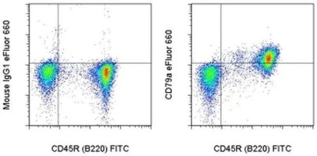
Applications Tested: This 24C2.5 antibody has been tested by intracellular staining and flow cytometric analysis of mouse splenocytes. This can be used at less than or equal to 1 µg per test. A test is defined as the amount (µg) of antibody that will stain a cell sample in a final volume of 100 µL. Cell number should be determined empirically but can range from 10^5 to 10^8 cells/test. It is recommended that the antibody be carefully titrated for optimal performance in the assay of interest.
eFluor® 660 is a replacement for Alexa Fluor® 647. eFluor® 660 emits at 659 nm and is excited with the red laser (633 nm). Please make sure that your instrument is capable of detecting this fluorochome.
Excitation: 633-647 nm; Emission: 668 nm; Laser: Red Laser.
Filtration: 0.2 µm post-manufacturing filtered.
For Research Use Only. Not for use in diagnostic procedures. Not for resale without express authorization.
CD79a Monoclonal Antibody (24C2.5), eFluor 660, eBioscience
PRODUCT DETAILS
Host: Mouse
Isotype: IgG1, kappa
Clonality: Monoclonal
Clone: 24C2.5
Format: eFluor 660
Reactivity: Ms
Application: Flow Cytometry
Tested Dilution: 1 µg/test
Concentration: 0.2 mg/mL
Storage: 4°C, store in dark, DO NOT FREEZE!
Formulation: PBS with 0.09% sodium azide; pH 7.2
Purification: Affinity chromatography
Data Sheet: TDS
Specific Information
Description: This 24C2.5 monoclonal antibody reacts with the intracellular tail of mouse CD79a (also known as Ig alpha or mb-1), a signaling component of the B cell receptor (BCR). CD79a heterodimerizes with CD79b (Ig beta, B29), and together with surface Ig, make up the BCR. Both CD79 subunits consist of a single extracellular Ig domain, a transmembrane domain, and an intracellular signaling domain. CD79a is expressed almost exclusively on B cells, as well as in most B lineage acute lymphoblastic leukemias. The cytoplasmic domain of CD79a contains immunoreceptor tyrosine-based activation motifs (ITAMs), which constitute the signal transducing portions of the BCR. Tyrosine phosphorylation of these ITAMs by Syk and Lyn initiates numerous signaling cascades, resulting in B cell activation, proliferation, and differentiation.
Applications Reported: This 24C2.5 antibody has been reported for use in intracellular staining followed by flow cytometric analysis.
Applications Tested: This 24C2.5 antibody has been tested by intracellular staining and flow cytometric analysis of mouse splenocytes. This can be used at less than or equal to 1 µg per test. A test is defined as the amount (µg) of antibody that will stain a cell sample in a final volume of 100 µL. Cell number should be determined empirically but can range from 10^5 to 10^8 cells/test. It is recommended that the antibody be carefully titrated for optimal performance in the assay of interest.
eFluor® 660 is a replacement for Alexa Fluor® 647. eFluor® 660 emits at 659 nm and is excited with the red laser (633 nm). Please make sure that your instrument is capable of detecting this fluorochome.
Excitation: 633-647 nm; Emission: 668 nm; Laser: Red Laser.
Filtration: 0.2 µm post-manufacturing filtered.
For Research Use Only. Not for use in diagnostic procedures. Not for resale without express authorization.
Product Information
Product Information
Shipping & Returns
Shipping & Returns
Description
PRODUCT DETAILS
Host: Mouse
Isotype: IgG1, kappa
Clonality: Monoclonal
Clone: 24C2.5
Format: eFluor 660
Reactivity: Ms
Application: Flow Cytometry
Tested Dilution: 1 µg/test
Concentration: 0.2 mg/mL
Storage: 4°C, store in dark, DO NOT FREEZE!
Formulation: PBS with 0.09% sodium azide; pH 7.2
Purification: Affinity chromatography
Data Sheet: TDS
Specific Information
Description: This 24C2.5 monoclonal antibody reacts with the intracellular tail of mouse CD79a (also known as Ig alpha or mb-1), a signaling component of the B cell receptor (BCR). CD79a heterodimerizes with CD79b (Ig beta, B29), and together with surface Ig, make up the BCR. Both CD79 subunits consist of a single extracellular Ig domain, a transmembrane domain, and an intracellular signaling domain. CD79a is expressed almost exclusively on B cells, as well as in most B lineage acute lymphoblastic leukemias. The cytoplasmic domain of CD79a contains immunoreceptor tyrosine-based activation motifs (ITAMs), which constitute the signal transducing portions of the BCR. Tyrosine phosphorylation of these ITAMs by Syk and Lyn initiates numerous signaling cascades, resulting in B cell activation, proliferation, and differentiation.
Applications Reported: This 24C2.5 antibody has been reported for use in intracellular staining followed by flow cytometric analysis.
Applications Tested: This 24C2.5 antibody has been tested by intracellular staining and flow cytometric analysis of mouse splenocytes. This can be used at less than or equal to 1 µg per test. A test is defined as the amount (µg) of antibody that will stain a cell sample in a final volume of 100 µL. Cell number should be determined empirically but can range from 10^5 to 10^8 cells/test. It is recommended that the antibody be carefully titrated for optimal performance in the assay of interest.
eFluor® 660 is a replacement for Alexa Fluor® 647. eFluor® 660 emits at 659 nm and is excited with the red laser (633 nm). Please make sure that your instrument is capable of detecting this fluorochome.
Excitation: 633-647 nm; Emission: 668 nm; Laser: Red Laser.
Filtration: 0.2 µm post-manufacturing filtered.
For Research Use Only. Not for use in diagnostic procedures. Not for resale without express authorization.


