CD68 Monoclonal Antibody (eBioY1/82A (Y1/82A)), eBioscience
PRODUCT DETAILS
Host: Mouse
Isotype: IgG2b, kappa
Clonality: Monoclonal
Clone: eBioY1/82A (Y1/82A)
Reactivity: Hu
Application: Flow Cytometry
Tested Dilution: 1 µg/test
Concentration: 0.5 mg/mL
Storage: 4°C
Formulation: PBS with 0.09% sodium azide; pH 7.2
Purification: Affinity chromatography
Data Sheet: TDS
Specific Information
Description: The eBioY1/82 antibody reacts with CD68 which belongs to the sialomucin family and is closely related to the family of lysosomal-associated membrane proteins (lamps) and scavenger receptor. CD68 is predominantly an intracellular protein, found mainly in the late endosomal compartment but can be detected in smaller amounts on the surface of cells of mainly myeloid derived cells; monocytes and macrophages, dendritic cells (and in some langerhan cells), neutrophils, basophils mast cells, myeloid progenitor cells, and a subset of CD34+ hemopoietic bone marrow progenitor cells. The function has not been fully elucidated but based on it's homolgy and structure suggests a role in antigen processing or presentation and protection of the lysosomal membrane from hydrolytic enzymes. Reports have also seen expression in activated T cells, and about 40% of peripheral blood B-lymphocytes and 50% of all B-ALL.
Staining with eBioY1/82A shows surface staining on B cells and a minor percentage of monocytes while intracellular staining results in about 50% of B cells and most monocytes.
Applications Reported: This eBioY1/82A (Y1/82A) antibody has been reported for use in flow cytometric analysis, and immunohistology staining of frozen tissue sections.
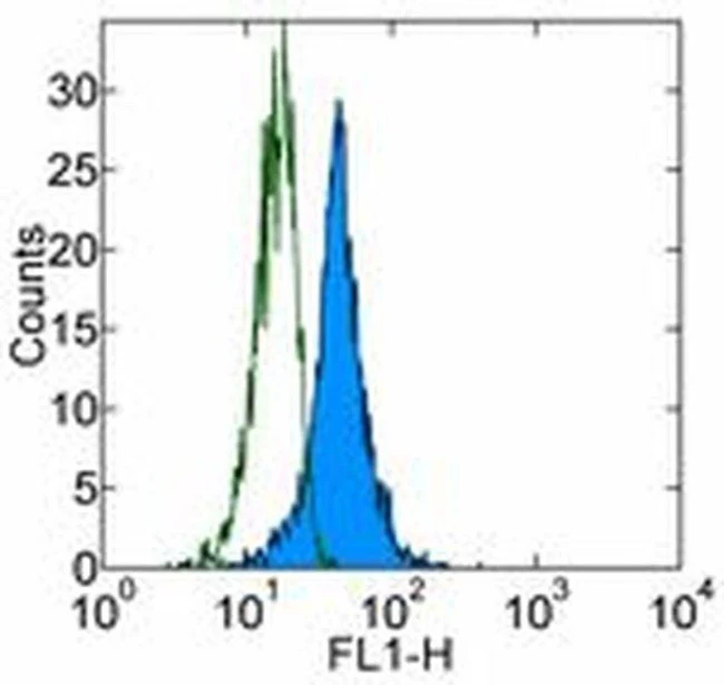
Applications Tested: This eBioY1/82A (Y1/82A) antibody has been tested by intracellular staining and flow cytometric analysis. This can be used at less than or equal to 1 µg per test. A test is defined as the amount (µg) of antibody that will stain a cell sample in a final volume of 100 µL. Cell number should be determined empirically but can range from 10^5 to 10^8 cells/test. It is recommended that the antibody be carefully titrated for optimal performance in the assay of interest.
Purity: Greater than 90%, as determined by SDS-PAGE.
Aggregation: Less than 10%, as determined by HPLC.
Filtration: 0.2 µm post-manufacturing filtered.
For Research Use Only. Not for use in diagnostic procedures. Not for resale without express authorization.
Original: $158.00
-70%$158.00
$47.40CD68 Monoclonal Antibody (eBioY1/82A (Y1/82A)), eBioscience
PRODUCT DETAILS
Host: Mouse
Isotype: IgG2b, kappa
Clonality: Monoclonal
Clone: eBioY1/82A (Y1/82A)
Reactivity: Hu
Application: Flow Cytometry
Tested Dilution: 1 µg/test
Concentration: 0.5 mg/mL
Storage: 4°C
Formulation: PBS with 0.09% sodium azide; pH 7.2
Purification: Affinity chromatography
Data Sheet: TDS
Specific Information
Description: The eBioY1/82 antibody reacts with CD68 which belongs to the sialomucin family and is closely related to the family of lysosomal-associated membrane proteins (lamps) and scavenger receptor. CD68 is predominantly an intracellular protein, found mainly in the late endosomal compartment but can be detected in smaller amounts on the surface of cells of mainly myeloid derived cells; monocytes and macrophages, dendritic cells (and in some langerhan cells), neutrophils, basophils mast cells, myeloid progenitor cells, and a subset of CD34+ hemopoietic bone marrow progenitor cells. The function has not been fully elucidated but based on it's homolgy and structure suggests a role in antigen processing or presentation and protection of the lysosomal membrane from hydrolytic enzymes. Reports have also seen expression in activated T cells, and about 40% of peripheral blood B-lymphocytes and 50% of all B-ALL.
Staining with eBioY1/82A shows surface staining on B cells and a minor percentage of monocytes while intracellular staining results in about 50% of B cells and most monocytes.
Applications Reported: This eBioY1/82A (Y1/82A) antibody has been reported for use in flow cytometric analysis, and immunohistology staining of frozen tissue sections.
Applications Tested: This eBioY1/82A (Y1/82A) antibody has been tested by intracellular staining and flow cytometric analysis. This can be used at less than or equal to 1 µg per test. A test is defined as the amount (µg) of antibody that will stain a cell sample in a final volume of 100 µL. Cell number should be determined empirically but can range from 10^5 to 10^8 cells/test. It is recommended that the antibody be carefully titrated for optimal performance in the assay of interest.
Purity: Greater than 90%, as determined by SDS-PAGE.
Aggregation: Less than 10%, as determined by HPLC.
Filtration: 0.2 µm post-manufacturing filtered.
For Research Use Only. Not for use in diagnostic procedures. Not for resale without express authorization.
Product Information
Product Information
Shipping & Returns
Shipping & Returns
Description
PRODUCT DETAILS
Host: Mouse
Isotype: IgG2b, kappa
Clonality: Monoclonal
Clone: eBioY1/82A (Y1/82A)
Reactivity: Hu
Application: Flow Cytometry
Tested Dilution: 1 µg/test
Concentration: 0.5 mg/mL
Storage: 4°C
Formulation: PBS with 0.09% sodium azide; pH 7.2
Purification: Affinity chromatography
Data Sheet: TDS
Specific Information
Description: The eBioY1/82 antibody reacts with CD68 which belongs to the sialomucin family and is closely related to the family of lysosomal-associated membrane proteins (lamps) and scavenger receptor. CD68 is predominantly an intracellular protein, found mainly in the late endosomal compartment but can be detected in smaller amounts on the surface of cells of mainly myeloid derived cells; monocytes and macrophages, dendritic cells (and in some langerhan cells), neutrophils, basophils mast cells, myeloid progenitor cells, and a subset of CD34+ hemopoietic bone marrow progenitor cells. The function has not been fully elucidated but based on it's homolgy and structure suggests a role in antigen processing or presentation and protection of the lysosomal membrane from hydrolytic enzymes. Reports have also seen expression in activated T cells, and about 40% of peripheral blood B-lymphocytes and 50% of all B-ALL.
Staining with eBioY1/82A shows surface staining on B cells and a minor percentage of monocytes while intracellular staining results in about 50% of B cells and most monocytes.
Applications Reported: This eBioY1/82A (Y1/82A) antibody has been reported for use in flow cytometric analysis, and immunohistology staining of frozen tissue sections.
Applications Tested: This eBioY1/82A (Y1/82A) antibody has been tested by intracellular staining and flow cytometric analysis. This can be used at less than or equal to 1 µg per test. A test is defined as the amount (µg) of antibody that will stain a cell sample in a final volume of 100 µL. Cell number should be determined empirically but can range from 10^5 to 10^8 cells/test. It is recommended that the antibody be carefully titrated for optimal performance in the assay of interest.
Purity: Greater than 90%, as determined by SDS-PAGE.
Aggregation: Less than 10%, as determined by HPLC.
Filtration: 0.2 µm post-manufacturing filtered.
For Research Use Only. Not for use in diagnostic procedures. Not for resale without express authorization.


