CD68 Monoclonal Antibody (eBioY1/82A (Y1/82A)), Alexa Fluor 647, eBioscience
PRODUCT DETAILS
Host: Mouse
Isotype: IgG2b, kappa
Clonality: Monoclonal
Clone: eBioY1/82A (Y1/82A)
Format: Alexa Fluor 647
Reactivity: Hu
Application: Flow Cytometry
Tested Dilution: 5 µL (0.125 µg)/test
Concentration: 5 μL/Test
Storage: 4°C, store in dark, DO NOT FREEZE!
Formulation: PBS with BSA and 0.09% sodium azide; pH 7.2
Purification: Affinity chromatography
Data Sheet: TDS
Specific Information
Description: The eBioY1/82A antibody reacts with CD68 which belongs to the sialomucin family and is closely related to the family of lysosomal-associated membrane proteins (LAMPs) and scavenger receptor. CD68 is predominantly an intracellular protein, found mainly in the late endosomal compartment but can be detected in smaller amounts on the surface of cells of mainly myeloid derived cells; monocytes and macrophages, dendritic cells (and in some Langerhans cells), neutrophils, basophils mast cells, myeloid progenitor cells, and a subset of CD34+ hemopoietic bone marrow progenitor cells. The function has not been fully elucidated but based on its homology and structure suggests a role in antigen processing or presentation and protection of the lysosomal membrane from hydrolytic enzymes. Reports have also seen expression in activated T cells, and about 40% of peripheral blood B-lymphocytes and 50% of all B-ALL.
Staining with eBioY1/82A shows surface staining on B cells and a minor percentage of monocytes while intracellular staining results in about 50% of B cells and most monocytes.
Applications Reported: This eBioY1/82A (Y1/82A) antibody has been reported for use in intracellular staining followed by flow cytometric analysis.
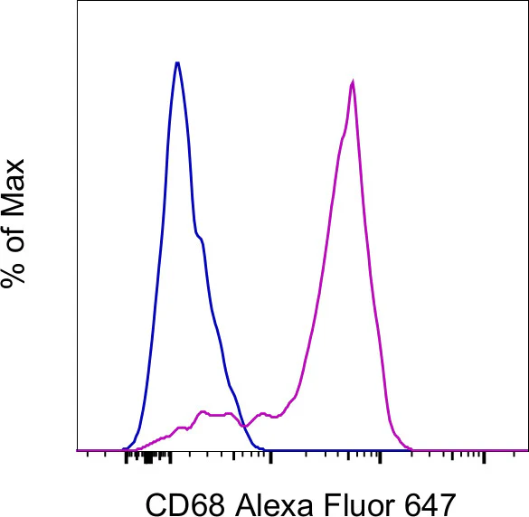
Applications Tested: This eBioY1/82A (Y1/82A) antibody has been pre-diluted and tested by intracellular staining followed by flow cytometric analysis of normal human peripheral blood cells using the Intracellular Fixation & Permeabilization Buffer Set (Product # 88-8824-00) and protocol. Please refer to "Staining Intracellular Antigens for Flow Cytometry, Protocol A: Two step protocol for intracellular (cytoplasmic) proteins" located at Flow Protocols. This may be used at 5 µL (0.125 µg) per test. A test is defined as the amount (µg) of antibody that will stain a cell sample in a final volume of 100 µL. Cell number should be determined empirically but can range from 10^5 to 10^8 cells/test.
Excitation: 633-647 nm; Emission: 668 nm; Laser: Red Laser.
For Research Use Only. Not for use in diagnostic procedures. Not for resale without express authorization.
Original: $359.00
-70%$359.00
$107.70CD68 Monoclonal Antibody (eBioY1/82A (Y1/82A)), Alexa Fluor 647, eBioscience
PRODUCT DETAILS
Host: Mouse
Isotype: IgG2b, kappa
Clonality: Monoclonal
Clone: eBioY1/82A (Y1/82A)
Format: Alexa Fluor 647
Reactivity: Hu
Application: Flow Cytometry
Tested Dilution: 5 µL (0.125 µg)/test
Concentration: 5 μL/Test
Storage: 4°C, store in dark, DO NOT FREEZE!
Formulation: PBS with BSA and 0.09% sodium azide; pH 7.2
Purification: Affinity chromatography
Data Sheet: TDS
Specific Information
Description: The eBioY1/82A antibody reacts with CD68 which belongs to the sialomucin family and is closely related to the family of lysosomal-associated membrane proteins (LAMPs) and scavenger receptor. CD68 is predominantly an intracellular protein, found mainly in the late endosomal compartment but can be detected in smaller amounts on the surface of cells of mainly myeloid derived cells; monocytes and macrophages, dendritic cells (and in some Langerhans cells), neutrophils, basophils mast cells, myeloid progenitor cells, and a subset of CD34+ hemopoietic bone marrow progenitor cells. The function has not been fully elucidated but based on its homology and structure suggests a role in antigen processing or presentation and protection of the lysosomal membrane from hydrolytic enzymes. Reports have also seen expression in activated T cells, and about 40% of peripheral blood B-lymphocytes and 50% of all B-ALL.
Staining with eBioY1/82A shows surface staining on B cells and a minor percentage of monocytes while intracellular staining results in about 50% of B cells and most monocytes.
Applications Reported: This eBioY1/82A (Y1/82A) antibody has been reported for use in intracellular staining followed by flow cytometric analysis.
Applications Tested: This eBioY1/82A (Y1/82A) antibody has been pre-diluted and tested by intracellular staining followed by flow cytometric analysis of normal human peripheral blood cells using the Intracellular Fixation & Permeabilization Buffer Set (Product # 88-8824-00) and protocol. Please refer to "Staining Intracellular Antigens for Flow Cytometry, Protocol A: Two step protocol for intracellular (cytoplasmic) proteins" located at Flow Protocols. This may be used at 5 µL (0.125 µg) per test. A test is defined as the amount (µg) of antibody that will stain a cell sample in a final volume of 100 µL. Cell number should be determined empirically but can range from 10^5 to 10^8 cells/test.
Excitation: 633-647 nm; Emission: 668 nm; Laser: Red Laser.
For Research Use Only. Not for use in diagnostic procedures. Not for resale without express authorization.
Product Information
Product Information
Shipping & Returns
Shipping & Returns
Description
PRODUCT DETAILS
Host: Mouse
Isotype: IgG2b, kappa
Clonality: Monoclonal
Clone: eBioY1/82A (Y1/82A)
Format: Alexa Fluor 647
Reactivity: Hu
Application: Flow Cytometry
Tested Dilution: 5 µL (0.125 µg)/test
Concentration: 5 μL/Test
Storage: 4°C, store in dark, DO NOT FREEZE!
Formulation: PBS with BSA and 0.09% sodium azide; pH 7.2
Purification: Affinity chromatography
Data Sheet: TDS
Specific Information
Description: The eBioY1/82A antibody reacts with CD68 which belongs to the sialomucin family and is closely related to the family of lysosomal-associated membrane proteins (LAMPs) and scavenger receptor. CD68 is predominantly an intracellular protein, found mainly in the late endosomal compartment but can be detected in smaller amounts on the surface of cells of mainly myeloid derived cells; monocytes and macrophages, dendritic cells (and in some Langerhans cells), neutrophils, basophils mast cells, myeloid progenitor cells, and a subset of CD34+ hemopoietic bone marrow progenitor cells. The function has not been fully elucidated but based on its homology and structure suggests a role in antigen processing or presentation and protection of the lysosomal membrane from hydrolytic enzymes. Reports have also seen expression in activated T cells, and about 40% of peripheral blood B-lymphocytes and 50% of all B-ALL.
Staining with eBioY1/82A shows surface staining on B cells and a minor percentage of monocytes while intracellular staining results in about 50% of B cells and most monocytes.
Applications Reported: This eBioY1/82A (Y1/82A) antibody has been reported for use in intracellular staining followed by flow cytometric analysis.
Applications Tested: This eBioY1/82A (Y1/82A) antibody has been pre-diluted and tested by intracellular staining followed by flow cytometric analysis of normal human peripheral blood cells using the Intracellular Fixation & Permeabilization Buffer Set (Product # 88-8824-00) and protocol. Please refer to "Staining Intracellular Antigens for Flow Cytometry, Protocol A: Two step protocol for intracellular (cytoplasmic) proteins" located at Flow Protocols. This may be used at 5 µL (0.125 µg) per test. A test is defined as the amount (µg) of antibody that will stain a cell sample in a final volume of 100 µL. Cell number should be determined empirically but can range from 10^5 to 10^8 cells/test.
Excitation: 633-647 nm; Emission: 668 nm; Laser: Red Laser.
For Research Use Only. Not for use in diagnostic procedures. Not for resale without express authorization.


