CD66a (CEACAM1) Monoclonal Antibody (CC1), PE, eBioscience
PRODUCT DETAILS
Host: Mouse
Isotype: IgG1, kappa
Clonality: Monoclonal
Clone: CC1
Format: PE
Reactivity: Ms
Application: Flow Cytometry
Tested Dilution: 0.06 µg/test
Concentration: 0.2 mg/mL
Storage: 4°C, store in dark, DO NOT FREEZE!
Formulation: PBS with 0.09% sodium azide; pH 7.2
Purification: Affinity chromatography
Data Sheet: TDS
Specific Information
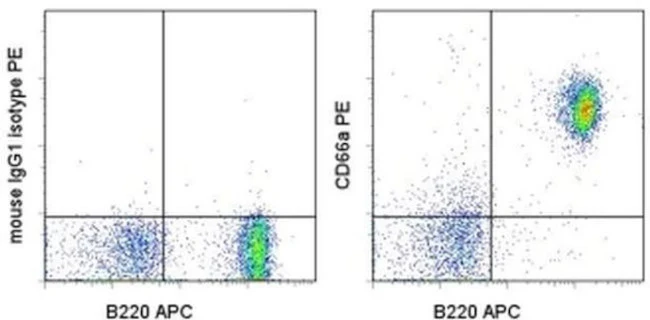
Description: The monoclonal antibody CC1 recognizes CD66a, also known as carcinoembryonic antigen-related cell adhesion molecule 1 (CEACAM1), biliary glycoprotein, and BPG. Expression of CD66a is found on brush borders, epithelial, and endothelial cells. In hematopoietic cells expression is found abundantly on B cells, as well as some NKs, monocytes, DCs, and granulocytes. Although low levels of mRNA have been identified in T cells in humans, resting mouse T lymphocytes are not reported to express CD66a, as confirmed by lack of staining with CC1 antibody. In humans, expression levels of CD66a have been used to identify malignancies. CD66a plays a key role as a regulator of BCR activation of B lymphocytes.
An alternate allele, CEACAM1b, is expressed in SJL mice; therefore, CC1 does not stain SJL tissue. The monoclonal CC1 has been shown to block viral infection and also enhance B cell proliferation when combined with IgM crosslinking.
Applications Reported: This CC1 antibody has been reported for use in flow cytometric analysis.
Applications Tested: This CC1 antibody has been tested by flow cytometric analysis of mouse splenocytes. This can be used at less than or equal to 0.06 µg per test. A test is defined as the amount (µg) of antibody that will stain a cell sample in a final volume of 100 µL. Cell number should be determined empirically but can range from 10^5 to 10^8 cells/test. It is recommended that the antibody be carefully titrated for optimal performance in the assay of interest.
Excitation: 488-561 nm; Emission: 578 nm; Laser: Blue Laser, Green Laser, Yellow-Green Laser.
Filtration: 0.2 µm post-manufacturing filtered.
For Research Use Only. Not for use in diagnostic procedures. Not for resale without express authorization.
CD66a (CEACAM1) Monoclonal Antibody (CC1), PE, eBioscience
PRODUCT DETAILS
Host: Mouse
Isotype: IgG1, kappa
Clonality: Monoclonal
Clone: CC1
Format: PE
Reactivity: Ms
Application: Flow Cytometry
Tested Dilution: 0.06 µg/test
Concentration: 0.2 mg/mL
Storage: 4°C, store in dark, DO NOT FREEZE!
Formulation: PBS with 0.09% sodium azide; pH 7.2
Purification: Affinity chromatography
Data Sheet: TDS
Specific Information
Description: The monoclonal antibody CC1 recognizes CD66a, also known as carcinoembryonic antigen-related cell adhesion molecule 1 (CEACAM1), biliary glycoprotein, and BPG. Expression of CD66a is found on brush borders, epithelial, and endothelial cells. In hematopoietic cells expression is found abundantly on B cells, as well as some NKs, monocytes, DCs, and granulocytes. Although low levels of mRNA have been identified in T cells in humans, resting mouse T lymphocytes are not reported to express CD66a, as confirmed by lack of staining with CC1 antibody. In humans, expression levels of CD66a have been used to identify malignancies. CD66a plays a key role as a regulator of BCR activation of B lymphocytes.
An alternate allele, CEACAM1b, is expressed in SJL mice; therefore, CC1 does not stain SJL tissue. The monoclonal CC1 has been shown to block viral infection and also enhance B cell proliferation when combined with IgM crosslinking.
Applications Reported: This CC1 antibody has been reported for use in flow cytometric analysis.
Applications Tested: This CC1 antibody has been tested by flow cytometric analysis of mouse splenocytes. This can be used at less than or equal to 0.06 µg per test. A test is defined as the amount (µg) of antibody that will stain a cell sample in a final volume of 100 µL. Cell number should be determined empirically but can range from 10^5 to 10^8 cells/test. It is recommended that the antibody be carefully titrated for optimal performance in the assay of interest.
Excitation: 488-561 nm; Emission: 578 nm; Laser: Blue Laser, Green Laser, Yellow-Green Laser.
Filtration: 0.2 µm post-manufacturing filtered.
For Research Use Only. Not for use in diagnostic procedures. Not for resale without express authorization.
Product Information
Product Information
Shipping & Returns
Shipping & Returns
Description
PRODUCT DETAILS
Host: Mouse
Isotype: IgG1, kappa
Clonality: Monoclonal
Clone: CC1
Format: PE
Reactivity: Ms
Application: Flow Cytometry
Tested Dilution: 0.06 µg/test
Concentration: 0.2 mg/mL
Storage: 4°C, store in dark, DO NOT FREEZE!
Formulation: PBS with 0.09% sodium azide; pH 7.2
Purification: Affinity chromatography
Data Sheet: TDS
Specific Information
Description: The monoclonal antibody CC1 recognizes CD66a, also known as carcinoembryonic antigen-related cell adhesion molecule 1 (CEACAM1), biliary glycoprotein, and BPG. Expression of CD66a is found on brush borders, epithelial, and endothelial cells. In hematopoietic cells expression is found abundantly on B cells, as well as some NKs, monocytes, DCs, and granulocytes. Although low levels of mRNA have been identified in T cells in humans, resting mouse T lymphocytes are not reported to express CD66a, as confirmed by lack of staining with CC1 antibody. In humans, expression levels of CD66a have been used to identify malignancies. CD66a plays a key role as a regulator of BCR activation of B lymphocytes.
An alternate allele, CEACAM1b, is expressed in SJL mice; therefore, CC1 does not stain SJL tissue. The monoclonal CC1 has been shown to block viral infection and also enhance B cell proliferation when combined with IgM crosslinking.
Applications Reported: This CC1 antibody has been reported for use in flow cytometric analysis.
Applications Tested: This CC1 antibody has been tested by flow cytometric analysis of mouse splenocytes. This can be used at less than or equal to 0.06 µg per test. A test is defined as the amount (µg) of antibody that will stain a cell sample in a final volume of 100 µL. Cell number should be determined empirically but can range from 10^5 to 10^8 cells/test. It is recommended that the antibody be carefully titrated for optimal performance in the assay of interest.
Excitation: 488-561 nm; Emission: 578 nm; Laser: Blue Laser, Green Laser, Yellow-Green Laser.
Filtration: 0.2 µm post-manufacturing filtered.
For Research Use Only. Not for use in diagnostic procedures. Not for resale without express authorization.


