CD56 (NCAM) Monoclonal Antibody (TULY56), PerCP-eFluor 710, eBioscience
PRODUCT DETAILS
Host: Mouse
Isotype: IgG1, kappa
Clonality: Monoclonal
Clone: TULY56
Format: PerCP-eFluor 710
Reactivity: Hu, Nhp, Rh
Application: Flow Cytometry
Tested Dilution: 5 µL (0.06 µg)/test
Concentration: 5 μL/Test
Storage: 4°C, store in dark, DO NOT FREEZE!
Formulation: PBS with BSA and 0.09% sodium azide
Purification: Affinity chromatography
Data Sheet: TDS
Specific Information
This TULY56 monoclonal antibody reacts with human CD56, also known as Neural Cell Adhesion Molecule (NCAM). CD56 is a highly glycosylated transmembrane molecule expressed by neurons and plays a role in the homotypic adhesion of neural cells. In the hematopoietic system, CD56 is expressed on NK cells and a subset of T cells referred to as NKT cells. Staining with TULY56 does not block binding of CMSSB, suggesting that the two antibodies recognize different epitopes. Additionally, TULY56 performs better after fixation and permeabilization than CMSSB.
The TULY56 monoclonal antibody crossreacts with Rhesus macaque.
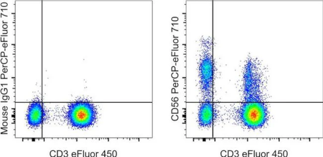
This TULY56 antibody has been pre-titrated and tested by flow cytometric analysis of normal human peripheral blood cells. This can be used at 5 µL (0.06 µg) per test. A test is defined as the amount (µg) of antibody that will stain a cell sample in a final volume of 100 µL. Cell number should be determined empirically but can range from 10^5 to 10^8 cells/test.
PerCP-eFluor® 710 emits at 710 nm and is excited with the blue laser (488 nm); it can be used in place of PerCP-Cyanine5.5. We recommend using a 710/50 bandpass filter, however, the 695/40 bandpass filter is an acceptable alternative. Please make sure that your instrument is capable of detecting this fluorochrome.
Light sensitivity: This tandem dye is sensitive to photo-induced oxidation. Please protect this vial and stained samples from light.
Fixation: Samples can be stored in IC Fixation Buffer (Product # 00-8222) (100 µL of cell sample + 100 µL of IC Fixation Buffer) or 1-step Fix/Lyse Solution (Product # 00-5333) for up to 3 days in the dark at 4°C with minimal impact on brightness and FRET efficiency/compensation. Some generalizations regarding fluorophore performance after fixation can be made, but clone specific performance should be determined empirically.
Filtration: 0.2 µm post-manufacturing filtered.
For Research Use Only. Not for use in diagnostic procedures. Not for resale without express authorization.
CD56 (NCAM) Monoclonal Antibody (TULY56), PerCP-eFluor 710, eBioscience
PRODUCT DETAILS
Host: Mouse
Isotype: IgG1, kappa
Clonality: Monoclonal
Clone: TULY56
Format: PerCP-eFluor 710
Reactivity: Hu, Nhp, Rh
Application: Flow Cytometry
Tested Dilution: 5 µL (0.06 µg)/test
Concentration: 5 μL/Test
Storage: 4°C, store in dark, DO NOT FREEZE!
Formulation: PBS with BSA and 0.09% sodium azide
Purification: Affinity chromatography
Data Sheet: TDS
Specific Information
This TULY56 monoclonal antibody reacts with human CD56, also known as Neural Cell Adhesion Molecule (NCAM). CD56 is a highly glycosylated transmembrane molecule expressed by neurons and plays a role in the homotypic adhesion of neural cells. In the hematopoietic system, CD56 is expressed on NK cells and a subset of T cells referred to as NKT cells. Staining with TULY56 does not block binding of CMSSB, suggesting that the two antibodies recognize different epitopes. Additionally, TULY56 performs better after fixation and permeabilization than CMSSB.
The TULY56 monoclonal antibody crossreacts with Rhesus macaque.
This TULY56 antibody has been pre-titrated and tested by flow cytometric analysis of normal human peripheral blood cells. This can be used at 5 µL (0.06 µg) per test. A test is defined as the amount (µg) of antibody that will stain a cell sample in a final volume of 100 µL. Cell number should be determined empirically but can range from 10^5 to 10^8 cells/test.
PerCP-eFluor® 710 emits at 710 nm and is excited with the blue laser (488 nm); it can be used in place of PerCP-Cyanine5.5. We recommend using a 710/50 bandpass filter, however, the 695/40 bandpass filter is an acceptable alternative. Please make sure that your instrument is capable of detecting this fluorochrome.
Light sensitivity: This tandem dye is sensitive to photo-induced oxidation. Please protect this vial and stained samples from light.
Fixation: Samples can be stored in IC Fixation Buffer (Product # 00-8222) (100 µL of cell sample + 100 µL of IC Fixation Buffer) or 1-step Fix/Lyse Solution (Product # 00-5333) for up to 3 days in the dark at 4°C with minimal impact on brightness and FRET efficiency/compensation. Some generalizations regarding fluorophore performance after fixation can be made, but clone specific performance should be determined empirically.
Filtration: 0.2 µm post-manufacturing filtered.
For Research Use Only. Not for use in diagnostic procedures. Not for resale without express authorization.
Product Information
Product Information
Shipping & Returns
Shipping & Returns
Description
PRODUCT DETAILS
Host: Mouse
Isotype: IgG1, kappa
Clonality: Monoclonal
Clone: TULY56
Format: PerCP-eFluor 710
Reactivity: Hu, Nhp, Rh
Application: Flow Cytometry
Tested Dilution: 5 µL (0.06 µg)/test
Concentration: 5 μL/Test
Storage: 4°C, store in dark, DO NOT FREEZE!
Formulation: PBS with BSA and 0.09% sodium azide
Purification: Affinity chromatography
Data Sheet: TDS
Specific Information
This TULY56 monoclonal antibody reacts with human CD56, also known as Neural Cell Adhesion Molecule (NCAM). CD56 is a highly glycosylated transmembrane molecule expressed by neurons and plays a role in the homotypic adhesion of neural cells. In the hematopoietic system, CD56 is expressed on NK cells and a subset of T cells referred to as NKT cells. Staining with TULY56 does not block binding of CMSSB, suggesting that the two antibodies recognize different epitopes. Additionally, TULY56 performs better after fixation and permeabilization than CMSSB.
The TULY56 monoclonal antibody crossreacts with Rhesus macaque.
This TULY56 antibody has been pre-titrated and tested by flow cytometric analysis of normal human peripheral blood cells. This can be used at 5 µL (0.06 µg) per test. A test is defined as the amount (µg) of antibody that will stain a cell sample in a final volume of 100 µL. Cell number should be determined empirically but can range from 10^5 to 10^8 cells/test.
PerCP-eFluor® 710 emits at 710 nm and is excited with the blue laser (488 nm); it can be used in place of PerCP-Cyanine5.5. We recommend using a 710/50 bandpass filter, however, the 695/40 bandpass filter is an acceptable alternative. Please make sure that your instrument is capable of detecting this fluorochrome.
Light sensitivity: This tandem dye is sensitive to photo-induced oxidation. Please protect this vial and stained samples from light.
Fixation: Samples can be stored in IC Fixation Buffer (Product # 00-8222) (100 µL of cell sample + 100 µL of IC Fixation Buffer) or 1-step Fix/Lyse Solution (Product # 00-5333) for up to 3 days in the dark at 4°C with minimal impact on brightness and FRET efficiency/compensation. Some generalizations regarding fluorophore performance after fixation can be made, but clone specific performance should be determined empirically.
Filtration: 0.2 µm post-manufacturing filtered.
For Research Use Only. Not for use in diagnostic procedures. Not for resale without express authorization.


