CD54 (ICAM-1) Monoclonal Antibody (eBioKAT-1 (KAT-1, KAT1)), eBioscience
PRODUCT DETAILS
Host: Rat
Isotype: IgG2a, kappa
Clonality: Monoclonal
Clone: eBioKAT-1 (KAT-1, KAT1)
Reactivity: Ms
Application: Flow Cytometry
Tested Dilution: 0.5 µg/test
Concentration: 0.5 mg/mL
Storage: 4°C
Formulation: PBS with 0.09% sodium azide; pH 7.2
Purification: Affinity chromatography
Data Sheet: TDS
Specific Information
Description: The eBioKAT-1 monoclonal antibody reacts with mouse CD54 (ICAM-1), which is a 95 kDa member of the immunoglobulin superfamily. CD54 is expressed at low levels on leukocytes and high endothelial venules, and expression increases in response to inflammatory cytokines. ICAM-1 binds to LFA-1 and this interaction is required for the transendothelial migration of T cells. ICAM-1-deficient mice are viable, however the migration of leukocytes to sites of inflammation is reduced leading to impaired immune and inflammatory responses. Based on the regulated expression of ICAM-1, it has been suggested that ICAM-1 may increase leukocyte extravasation at sites of inflammation, whereas the constitutively high expression of ICAM-2 mediates leukocyte traffic into non-inflamed tissue. The eBioKAT-1 monoclonal antibody recognizes a different epitope than the YN1/1.7.4 monoclonal antibody.
Applications Reported: This eBioKAT-1 (KAT-1, KAT1) antibody has been reported for use in flow cytometric analysis, immunoprecipitation, immunohistology staining of frozen tissue sections, and immunohistology staining of paraffin embedded tissue sections. (Please use Functional Grade purified eBioKAT-1 (KAT-1, KAT1), Product # 16-0542, in functional assays).
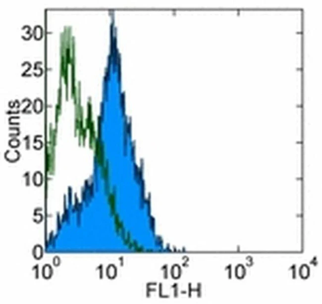
Applications Tested: This eBioKAT-1 (KAT-1, KAT1) antibody has been tested by flow cytometric analysis of mouse splenocytes. This can be used at less than or equal to 0.5 µg per test. A test is defined as the amount (µg) of antibody that will stain a cell sample in a final volume of 100 µL. Cell number should be determined empirically but can range from 10^5 to 10^8 cells/test. It is recommended that the antibody be carefully titrated for optimal performance in the assay of interest.
Purity: Greater than 90%, as determined by SDS-PAGE.
Aggregation: Less than 10%, as determined by HPLC.
Filtration: 0.2 µm post-manufacturing filtered.
For Research Use Only. Not for use in diagnostic procedures. Not for resale without express authorization.
Original: $142.00
-70%$142.00
$42.60CD54 (ICAM-1) Monoclonal Antibody (eBioKAT-1 (KAT-1, KAT1)), eBioscience
PRODUCT DETAILS
Host: Rat
Isotype: IgG2a, kappa
Clonality: Monoclonal
Clone: eBioKAT-1 (KAT-1, KAT1)
Reactivity: Ms
Application: Flow Cytometry
Tested Dilution: 0.5 µg/test
Concentration: 0.5 mg/mL
Storage: 4°C
Formulation: PBS with 0.09% sodium azide; pH 7.2
Purification: Affinity chromatography
Data Sheet: TDS
Specific Information
Description: The eBioKAT-1 monoclonal antibody reacts with mouse CD54 (ICAM-1), which is a 95 kDa member of the immunoglobulin superfamily. CD54 is expressed at low levels on leukocytes and high endothelial venules, and expression increases in response to inflammatory cytokines. ICAM-1 binds to LFA-1 and this interaction is required for the transendothelial migration of T cells. ICAM-1-deficient mice are viable, however the migration of leukocytes to sites of inflammation is reduced leading to impaired immune and inflammatory responses. Based on the regulated expression of ICAM-1, it has been suggested that ICAM-1 may increase leukocyte extravasation at sites of inflammation, whereas the constitutively high expression of ICAM-2 mediates leukocyte traffic into non-inflamed tissue. The eBioKAT-1 monoclonal antibody recognizes a different epitope than the YN1/1.7.4 monoclonal antibody.
Applications Reported: This eBioKAT-1 (KAT-1, KAT1) antibody has been reported for use in flow cytometric analysis, immunoprecipitation, immunohistology staining of frozen tissue sections, and immunohistology staining of paraffin embedded tissue sections. (Please use Functional Grade purified eBioKAT-1 (KAT-1, KAT1), Product # 16-0542, in functional assays).
Applications Tested: This eBioKAT-1 (KAT-1, KAT1) antibody has been tested by flow cytometric analysis of mouse splenocytes. This can be used at less than or equal to 0.5 µg per test. A test is defined as the amount (µg) of antibody that will stain a cell sample in a final volume of 100 µL. Cell number should be determined empirically but can range from 10^5 to 10^8 cells/test. It is recommended that the antibody be carefully titrated for optimal performance in the assay of interest.
Purity: Greater than 90%, as determined by SDS-PAGE.
Aggregation: Less than 10%, as determined by HPLC.
Filtration: 0.2 µm post-manufacturing filtered.
For Research Use Only. Not for use in diagnostic procedures. Not for resale without express authorization.
Product Information
Product Information
Shipping & Returns
Shipping & Returns
Description
PRODUCT DETAILS
Host: Rat
Isotype: IgG2a, kappa
Clonality: Monoclonal
Clone: eBioKAT-1 (KAT-1, KAT1)
Reactivity: Ms
Application: Flow Cytometry
Tested Dilution: 0.5 µg/test
Concentration: 0.5 mg/mL
Storage: 4°C
Formulation: PBS with 0.09% sodium azide; pH 7.2
Purification: Affinity chromatography
Data Sheet: TDS
Specific Information
Description: The eBioKAT-1 monoclonal antibody reacts with mouse CD54 (ICAM-1), which is a 95 kDa member of the immunoglobulin superfamily. CD54 is expressed at low levels on leukocytes and high endothelial venules, and expression increases in response to inflammatory cytokines. ICAM-1 binds to LFA-1 and this interaction is required for the transendothelial migration of T cells. ICAM-1-deficient mice are viable, however the migration of leukocytes to sites of inflammation is reduced leading to impaired immune and inflammatory responses. Based on the regulated expression of ICAM-1, it has been suggested that ICAM-1 may increase leukocyte extravasation at sites of inflammation, whereas the constitutively high expression of ICAM-2 mediates leukocyte traffic into non-inflamed tissue. The eBioKAT-1 monoclonal antibody recognizes a different epitope than the YN1/1.7.4 monoclonal antibody.
Applications Reported: This eBioKAT-1 (KAT-1, KAT1) antibody has been reported for use in flow cytometric analysis, immunoprecipitation, immunohistology staining of frozen tissue sections, and immunohistology staining of paraffin embedded tissue sections. (Please use Functional Grade purified eBioKAT-1 (KAT-1, KAT1), Product # 16-0542, in functional assays).
Applications Tested: This eBioKAT-1 (KAT-1, KAT1) antibody has been tested by flow cytometric analysis of mouse splenocytes. This can be used at less than or equal to 0.5 µg per test. A test is defined as the amount (µg) of antibody that will stain a cell sample in a final volume of 100 µL. Cell number should be determined empirically but can range from 10^5 to 10^8 cells/test. It is recommended that the antibody be carefully titrated for optimal performance in the assay of interest.
Purity: Greater than 90%, as determined by SDS-PAGE.
Aggregation: Less than 10%, as determined by HPLC.
Filtration: 0.2 µm post-manufacturing filtered.
For Research Use Only. Not for use in diagnostic procedures. Not for resale without express authorization.


