CD366 (TIM3) Monoclonal Antibody (RMT3-23), Super Bright™ 702, eBioscience™
PRODUCT DETAILS
Host: Rat
Isotype: IgG2a, kappa
Clonality: Monoclonal
Clone: RMT3-23
Format: Super Bright™ 702
Reactivity: Mouse
Application: Flow Cytometry
Tested Dilution: 0.5 µg/test
Concentration: 0.2 mg/mL
Storage: 4° C, store in dark, DO NOT FREEZE!
Formulation: PBS, pH 7.2, containing 0.09% sodium azide
Purification: Affinity chromatography
Data Sheet: TDS
Specific Information
Description: The RMT3-23 monoclonal antibody reacts with mouse CD366 (TIM3), a Th1-specific cell surface protein. The RMT3-23 antibody reacts with CD366 protein expressed by both BALB/c and C57BL/6 strains of mice. CD366, a type I transmembrane protein, contains an immunoglobulin and a mucin-like domain in its extracellular portion and a tyrosine phosphorylation motif in its cytoplasmic portion. CD366 is expressed selectively by differentiated CD4^+Th1 and CD8^+Tc1 cells, but is absent on CD4^+Th2 and CD8^+Tc2 cells. Other hematopoietic cell types, including naive T cells, B cells, macrophages and dendritic cells, do not express CD366, at least at the protein level. CD366 expression is upregulated at a late stage of T cell differentiation on Th1 cells after 3 rounds of in vitro polarization suggesting a role for this molecule in the transport or effector function of Th1 cells rather than a contribution to T cell differentiation. In an experimental autoimmune encephalomyelitis (EAE) model, CD366 was shown to be expressed on most CD4^+ and CD8^+ T cells in the central nervous system at the onset of clinical signs of disease, while less than 2% of CD4^+ cells in the periphery expressed CD366 after immunization.
Applications Reported: This RMT3-23 antibody has been reported for use in flow cytometric analysis.
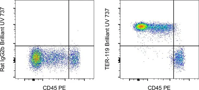
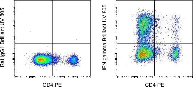
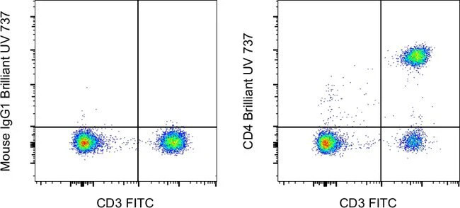
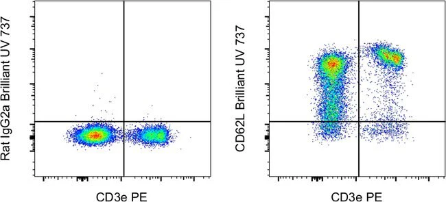
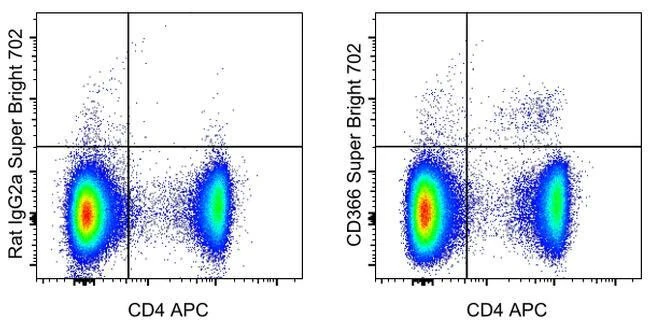
Applications Tested: This RMT3-23 antibody has been tested by flow cytometric analysis of mouse splenocytes. This may be used at less than or equal to 0.5 µg per test. A test is defined as the amount (µg) of antibody that will stain a cell sample in a final volume of 100 µL. Cell number should be determined empirically but can range from 10^5 to 10^8 cells/test. It is recommended that the antibody be carefully titrated for optimal performance in the assay of interest.
Super Bright 702 is a tandem dye that can be excited with the violet laser line (405 nm) and emits at 702 nm. We recommend using a 710/50 bandpass filter. Please make sure that your instrument is capable of detecting this fluorochrome.
When using two or more Super Bright dye-conjugated antibodies in a staining panel, it is recommended to use Super Bright Complete Staining Buffer (Product # SB-4401) to minimize any non-specific polymer interactions. Please refer to the datasheet for Super Bright Staining Buffer for more information.
Light sensitivity: This tandem dye is sensitive to photo-induced oxidation. Please protect this vial and stained samples from light.
Fixation: Samples can be stored in IC Fixation Buffer (Product # 00-8222) (100 µL of cell sample + 100 µL of IC Fixation Buffer) or 1-step Fix/Lyse Solution (Product # 00-5333) for up to 3 days in the dark at 4°C with minimal impact on brightness and FRET efficiency/compensation. Some generalizations regarding fluorophore performance after fixation can be made, but clone specific performance should be determined empirically.
Excitation: 405 nm; Emission: 702 nm; Laser: Violet Laser
Super Bright Polymer Dyes are sold under license from Becton, Dickinson and Company.
For Research Use Only. Not for use in diagnostic procedures. Not for resale without express authorization.
CD366 (TIM3) Monoclonal Antibody (RMT3-23), Super Bright™ 702, eBioscience™
PRODUCT DETAILS
Host: Rat
Isotype: IgG2a, kappa
Clonality: Monoclonal
Clone: RMT3-23
Format: Super Bright™ 702
Reactivity: Mouse
Application: Flow Cytometry
Tested Dilution: 0.5 µg/test
Concentration: 0.2 mg/mL
Storage: 4° C, store in dark, DO NOT FREEZE!
Formulation: PBS, pH 7.2, containing 0.09% sodium azide
Purification: Affinity chromatography
Data Sheet: TDS
Specific Information
Description: The RMT3-23 monoclonal antibody reacts with mouse CD366 (TIM3), a Th1-specific cell surface protein. The RMT3-23 antibody reacts with CD366 protein expressed by both BALB/c and C57BL/6 strains of mice. CD366, a type I transmembrane protein, contains an immunoglobulin and a mucin-like domain in its extracellular portion and a tyrosine phosphorylation motif in its cytoplasmic portion. CD366 is expressed selectively by differentiated CD4^+Th1 and CD8^+Tc1 cells, but is absent on CD4^+Th2 and CD8^+Tc2 cells. Other hematopoietic cell types, including naive T cells, B cells, macrophages and dendritic cells, do not express CD366, at least at the protein level. CD366 expression is upregulated at a late stage of T cell differentiation on Th1 cells after 3 rounds of in vitro polarization suggesting a role for this molecule in the transport or effector function of Th1 cells rather than a contribution to T cell differentiation. In an experimental autoimmune encephalomyelitis (EAE) model, CD366 was shown to be expressed on most CD4^+ and CD8^+ T cells in the central nervous system at the onset of clinical signs of disease, while less than 2% of CD4^+ cells in the periphery expressed CD366 after immunization.
Applications Reported: This RMT3-23 antibody has been reported for use in flow cytometric analysis.
Applications Tested: This RMT3-23 antibody has been tested by flow cytometric analysis of mouse splenocytes. This may be used at less than or equal to 0.5 µg per test. A test is defined as the amount (µg) of antibody that will stain a cell sample in a final volume of 100 µL. Cell number should be determined empirically but can range from 10^5 to 10^8 cells/test. It is recommended that the antibody be carefully titrated for optimal performance in the assay of interest.
Super Bright 702 is a tandem dye that can be excited with the violet laser line (405 nm) and emits at 702 nm. We recommend using a 710/50 bandpass filter. Please make sure that your instrument is capable of detecting this fluorochrome.
When using two or more Super Bright dye-conjugated antibodies in a staining panel, it is recommended to use Super Bright Complete Staining Buffer (Product # SB-4401) to minimize any non-specific polymer interactions. Please refer to the datasheet for Super Bright Staining Buffer for more information.
Light sensitivity: This tandem dye is sensitive to photo-induced oxidation. Please protect this vial and stained samples from light.
Fixation: Samples can be stored in IC Fixation Buffer (Product # 00-8222) (100 µL of cell sample + 100 µL of IC Fixation Buffer) or 1-step Fix/Lyse Solution (Product # 00-5333) for up to 3 days in the dark at 4°C with minimal impact on brightness and FRET efficiency/compensation. Some generalizations regarding fluorophore performance after fixation can be made, but clone specific performance should be determined empirically.
Excitation: 405 nm; Emission: 702 nm; Laser: Violet Laser
Super Bright Polymer Dyes are sold under license from Becton, Dickinson and Company.
For Research Use Only. Not for use in diagnostic procedures. Not for resale without express authorization.
Product Information
Product Information
Shipping & Returns
Shipping & Returns
Description
PRODUCT DETAILS
Host: Rat
Isotype: IgG2a, kappa
Clonality: Monoclonal
Clone: RMT3-23
Format: Super Bright™ 702
Reactivity: Mouse
Application: Flow Cytometry
Tested Dilution: 0.5 µg/test
Concentration: 0.2 mg/mL
Storage: 4° C, store in dark, DO NOT FREEZE!
Formulation: PBS, pH 7.2, containing 0.09% sodium azide
Purification: Affinity chromatography
Data Sheet: TDS
Specific Information
Description: The RMT3-23 monoclonal antibody reacts with mouse CD366 (TIM3), a Th1-specific cell surface protein. The RMT3-23 antibody reacts with CD366 protein expressed by both BALB/c and C57BL/6 strains of mice. CD366, a type I transmembrane protein, contains an immunoglobulin and a mucin-like domain in its extracellular portion and a tyrosine phosphorylation motif in its cytoplasmic portion. CD366 is expressed selectively by differentiated CD4^+Th1 and CD8^+Tc1 cells, but is absent on CD4^+Th2 and CD8^+Tc2 cells. Other hematopoietic cell types, including naive T cells, B cells, macrophages and dendritic cells, do not express CD366, at least at the protein level. CD366 expression is upregulated at a late stage of T cell differentiation on Th1 cells after 3 rounds of in vitro polarization suggesting a role for this molecule in the transport or effector function of Th1 cells rather than a contribution to T cell differentiation. In an experimental autoimmune encephalomyelitis (EAE) model, CD366 was shown to be expressed on most CD4^+ and CD8^+ T cells in the central nervous system at the onset of clinical signs of disease, while less than 2% of CD4^+ cells in the periphery expressed CD366 after immunization.
Applications Reported: This RMT3-23 antibody has been reported for use in flow cytometric analysis.
Applications Tested: This RMT3-23 antibody has been tested by flow cytometric analysis of mouse splenocytes. This may be used at less than or equal to 0.5 µg per test. A test is defined as the amount (µg) of antibody that will stain a cell sample in a final volume of 100 µL. Cell number should be determined empirically but can range from 10^5 to 10^8 cells/test. It is recommended that the antibody be carefully titrated for optimal performance in the assay of interest.
Super Bright 702 is a tandem dye that can be excited with the violet laser line (405 nm) and emits at 702 nm. We recommend using a 710/50 bandpass filter. Please make sure that your instrument is capable of detecting this fluorochrome.
When using two or more Super Bright dye-conjugated antibodies in a staining panel, it is recommended to use Super Bright Complete Staining Buffer (Product # SB-4401) to minimize any non-specific polymer interactions. Please refer to the datasheet for Super Bright Staining Buffer for more information.
Light sensitivity: This tandem dye is sensitive to photo-induced oxidation. Please protect this vial and stained samples from light.
Fixation: Samples can be stored in IC Fixation Buffer (Product # 00-8222) (100 µL of cell sample + 100 µL of IC Fixation Buffer) or 1-step Fix/Lyse Solution (Product # 00-5333) for up to 3 days in the dark at 4°C with minimal impact on brightness and FRET efficiency/compensation. Some generalizations regarding fluorophore performance after fixation can be made, but clone specific performance should be determined empirically.
Excitation: 405 nm; Emission: 702 nm; Laser: Violet Laser
Super Bright Polymer Dyes are sold under license from Becton, Dickinson and Company.
For Research Use Only. Not for use in diagnostic procedures. Not for resale without express authorization.